Lupine Publishers Group
Lupine Publishers
Menu
ISSN: 2637-4544
Research Article(ISSN: 2637-4544) 
Preeclampsia Predictability Tools using Trace Metal Screening and Angiogenic Markers is Clinically Valuable Volume 3 - Issue 5
Ayman El-Dorf1* and Maha M Hagras2
- 1Obstetrics and Gynecology Department, Faculty of Medicine, Tanta University, Tanta, Egypt
- 2Clinical Pathology Department, Faculty of Medicine, Tanta University, Tanta, Egypt
Received:September 03, 2019; Published: September 30, 2019
Corresponding author: Ayman El Dorf, Obstetrics and Gynecology Department, Faculty of Medicine, Tanta University, Tanta, Egypt
DOI: 10.32474/IGWHC.2019.03.000175
Abstract
Background: Preeclampsia at molecular and cellular levels was observed as a disease of placentation that is affected by interaction of various factors that could trigger the disease pathological course and behavior. Prenatal environmental metals exposure is considered a cornerstone factor that could trigger the clinical presentation of preeclampsia due to toxic effects of some trace metals, besides the deficiency of some trace elements is considered a crucial issue in cell apoptosis and remodeling is one of the characteristic features of trophoblastic invasion.
Aim: To investigate the predictability value of trace metal screening in predictability of preeclampsia clinical development.
Methodology: The current research clinical trial is prospective in manner that recruited 420 research study subjects from January 2017 till April 2019 ,inclusive research criteria were singleton gestations with no congenital fetal anomalies all recruited research study subjects had full clinical history taking and examination during antenatal visits urinary samples obtained during 10th till 15th gestational weeks were assessed for the trace metal profile investigated and angiogenic markers. Urine samples were collected at 10th till 15th gestational weeks and stored at − 80 °C until analysis after delivery, preeclampsia diagnosis and diagnosis date were abstracted from medical records. Preeclampsia was clinically defined as elevated maternal blood pressure (>140mmHg systolic and/or >90mmHg diastolic) and proteinuria (>300 mg/24 h or a protein/creatinine ratio >0.20) after 20 gestational weeks.
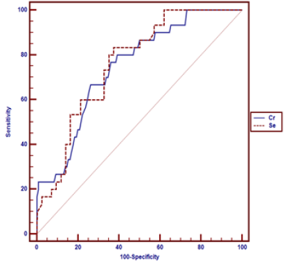
Results: Cr chromium at Cut off point >0.92 have AUC= 0.747, statistical Sensitivity= 80.0, statistical Specificity=60.8, PPV=13.6, NPV =97.5, Se selenium at cutoff point ≤ 35.4 have AUC= 0.759, statistical Sensitivity=83.3, statistical Specificity=62.6, PPV=14.6, NPV =98.
Conclusion: The current study provokes the clinical value for trace metal screening in detectability of preeclamptic risk of development, however future research efforts are requiring correlating the clinical risk of disease development according to the environmental trace metal levels.
Introduction
Preeclampsia is a global reproductive disease that is characterized by elevated blood pressure levels and proteinuria after 20 gestational weeks, various theories were proposed by researchers that leaves preeclampsia pathophysiology a complex disease as regard the pathophysiological basis [1-3]. Preeclampsia at molecular and cellular levels was observed as a disease of placentation that is affected by interaction of various factors that could trigger the disease pathological course and behavior. A growing interest all over the world is to reveal any biomarkers that could early detect the disease development in a manner that could improve the clinical management of the disease reducing the morbidity and mortality issues [4-6].
Disordered physiological process of spiral artery remodeling, have raised the interest of using angiogenic biomarkers for predictability of preeclampsia risk, soluble fms-like tyrosine kinase-1 and placental growth factor are prominent angiogenic biomarkers that could reflect the effectiveness and adequacy of placental angiogenesis process and are considered family members of vascular endothelial growth factors .integration of both biomarkers particularly when used in the form of ratio was observed by prior research groups to be highly effective in detectability of pre-eclamptic issues arousal [7-9]. Prenatal environmental metals exposure is considered a cornerstone factor that could trigger the clinical presentation of preeclampsia due to toxic effects of some trace metals, besides the deficiency of some trace elements is considered a crucial issue in cell apoptosis and remodeling is one of the characteristic features of trophoblastic invasion. cadmium (Cd) and lead (Pb), have been observed by various researchers could cause teratogenic impacts on the fetal developmental process particularly within the first gestational trimester besides their well demonstrated raised risk of preeclampsia development [10-12].
On the other hand, it was demonstrated in an interesting manner that the elevated, exposure to essential metals, e.g. selenium (Se) and zinc (Zn), within the safe recommended range values is correlated to higher fertility potential and reduced preeclampsia clinical risk. Those research findings could be justified by the fact that toxic metals could trigger oxidative stress within the placental environment besides causing impaired trophoblastic invasion .another issue of concern that have been displayed from prior research groups is that the toxic metals could provoke immunological abnormalities that would consequently affect the trophoblastic invasion process that relies on complex immunological interactions that safe guard the fetus from allograft process of rejection and it is well known that the affection of immunological adaptive process integrity is a pathophysiological triggering issue for preeclampsia development [13-15]. Essential metals are considered the molecular factors that reduce the oxidative stress that would cause cellular damage therefore their existence in adequate physiological levels would prevent oxidative stress pathological sequale that is well observed in preeclampsia and infertility cases in numerous research efforts [16-18].
Methodology
The current research clinical trial is prospective in manner that recruited 420 research study subjects from January 2017 till April 2019 ,inclusive research criteria were singleton gestations with no congenital fetal anomalies had full clinical history taking and examination during antenatal visits. urinary samples and venous blood samples obtained during 10th till 15th gestational weeks. urinary samples were assessed for the trace metal profile investigated and venous blood samples were assessed for angiogenic markers, exclusive research criteria were cases with multiple pregnancies and fetal anomalies observed during sonographic scanning during last first trimetric scanning, or the mid trimetric full anomaly scanning during antenatal visits all cases were followed up for medical disease development such as DM and preeclampsia development according to preeclampsia development the cases were categorized into two research groups preeclampsia and non-preeclampsia research groups the research data of both groups were statically analyzed in a comparative a manner in correlations to the levels of trace metals investigated and angiogenic markers. Urine samples were collected at 10th till 15th gestational weeks and stored at − 80 °C until analysis after delivery, preeclampsia diagnosis and diagnosis date were abstracted from medical records. Preeclampsia was clinically defined as elevated maternal blood pressure (> 140mmHg systolic and/or > 90mmHg diastolic) and proteinuria (> 300 mg/24 h or a protein/creatinine ratio > 0.20) after 20 gestational weeks.
Urinary Trace Metals Analysis
Twenty four hour urine samples were collected at 10th till 15th gestational weeks and stored at − 80 °C in airtight polypropylene containers until analysis after delivery. Trace metals were analyzed using inductively coupled plasma mass spectrometry (ICP-MS), The metals analyzed included arsenic (As), barium (Ba), beryllium (Be), Cd, copper (Cu), chromium (Cr), mercury (Hg), manganese (Mn), molybdenum (Mo), nickel (Ni), Pb, Se, tin (Sn), thallium (Tl), uranium (U), tungsten (W), and Zn.
Plasma Biomarkers of Angiogenesis
Venous blood samples(5 ml) obtained during 10th till 15th gestational weeks under complete aseptic conditions, allowed to clot and then centrifuged at 3000 rpm for 10 minutes to separate serum that was collected in sterile Eppindorff tube and stored at -10°C till be assayed for circulating maternal sFlt-1 and PlGF Assay of circulating maternal sFlt-1 and PlGF During 10th till 15th gestational trimester using ARCHITECT immunoassays (Abbot Laboratory, Abbott Park, IL, USA). Specifically, unbound PlGF concentrations from 1 to 1500 pg/mL have been assayed and total (unbound and bound) sFlt-1 concentrations from 0.10–150 ng/mL were measured. The ratio of sFlt-1 to PlGF was also calculated for analysis the combined intra- and intraassay coefficients of variation were < 7% for both PlGF and sFlt-1.
Statistical Analysis
Data were collected, revised and performed using SPSS version 23. The data were presented as numbers, percentages, mean, standard deviations and median with inter-quartile range (IQR) and the differences between the two groups were assessed using T-tests, Mann-Whitney test, Chi-square tests, or Fisher’s exact tests, when appropriate. Logistic regression analysis for urinary metals as predictors of preeclampsia adjusted for specific gravity, smoking during pregnancy, educational attainment, infant sex, ART, calcium supplementation, pre-pregnancy BMI, and gestational age at study visit to assess. Also, Receiver operating characteristic curve was used to assess the best cut off point with its sensitivity, specificity, positive predictive value (PPV), negative predictive value (NPV) and area under curve (AUC) of the significant urinary metals associated to preeclampsia. The confidence interval was set to 95% and the margin of error accepted was set to 5%. So, the p-value was considered significant at the level of < 0.05.
Results
Table 1 reveals and displays the Demographic characteristics of the investigated research groups in which there was no statistical significant difference as regards maternal age, education, smoking and alcohol consumption during pregnancy, parity, gestational DM, Usage of Multivitamins, Calcium Supplements, Iron Supplement During Pregnancy, Maternal sFlt-1Expression (p values=0.453, 0.295 ,0.407, 0.703, 0.869, 0.407, 0.809,0.063, 0.681, 0.410 consecutively )whereas there is statically significant difference between both research groups as regards Pre-pregnancy BMI (kg/m2), Previous Preeclampsia Diagnosis, Use of ART, Chronic Hypertension, Infant Sex (males), Maternal PLGF Expression ,Maternal sFlt-1/PLGF Ratio (p values <0.001, <0.001, <0.001, <0.001, 0.004, <0.001, 0.004 consecutively).
Table 1: Comparison of maternal characteristics and outcomes for the two study groups. Data were presented as mean and standard deviations, median (IQR) and numbers and percentages •: Independent t-test; ≠: Mann-Whitney test; *: Chi-square test.
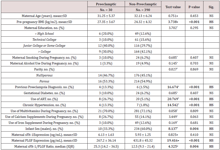
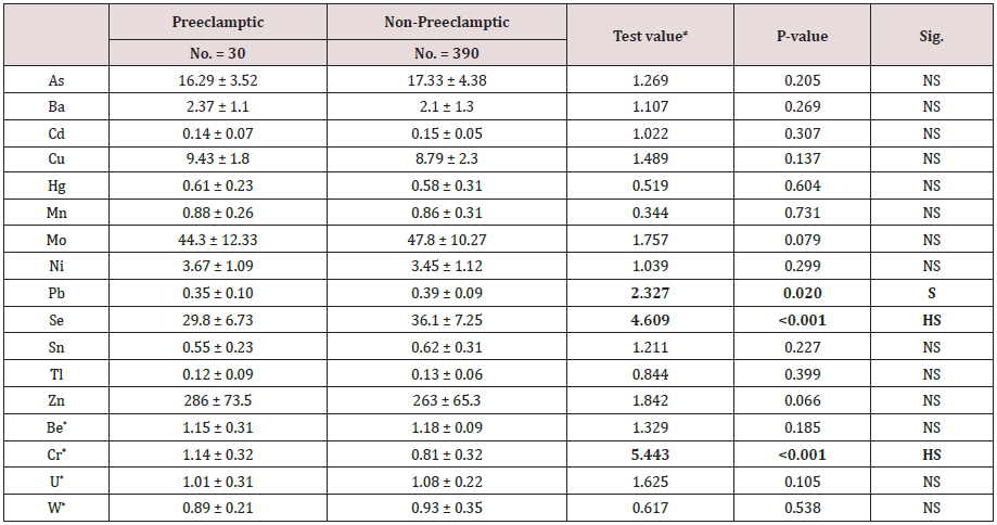
Table 2 reveals and displays the comparative analysis between preeclamptic and non -preeclamptic research groups as regards trace metals from urine samples (μg/L) Showing no statistical significant difference concerning As arsenic, Ba barium, Cd cadmium, Cu copper, Hg mercury, Mn manganese, Mo molybdenum, Ni nickel, Sn tin, Tl thallium, Zn zinc, Be beryllium, U uranium, W tungsten(p values=0.205, 0.269, 0.307, 0.137, 0.604, 0.731, 0.073, 0.299, 0.227, 0.399, 0.066, 0.185, 0.105, 0.538 consecutively) whereas the levels of Pb lead, Se selenium , Cr chromium whereas statistically significantly different between both research groups (p values =0.020, <0.001, <0.001 consecutively). Table 3 reveals and displays the Adjusted logistic regression analysis for urinary metals as predictors of preeclampsia revealing that there was no statistical significance as regards As arsenic, Ba barium, Cd cadmium, Cu copper, Hg mercury, Mn manganese, Mo molybdenum, Ni nickel, Pb lead, Sn tin, Tl thallium, Zn zinc, Be beryllium, U uranium, W tungsten (p values= 0.125, 0.673, 0.713, 0.512, 0.670, 0.295, 0.091, 0.733, 0.356, 0.654,0.654, 0.513, 0.812, 0.903, 0.511 consecutively) whereas there was stoical significance concerning, Se selenium, Cr chromium (p values=0.032, 0.002 consecutively).
Table 3: Relation between adverse neonatal outcome and fetal urinary production rate, adjusted for gestational age. FUPR: Fetal Urinary Production Rate; CSF: Cerebrospinal Fluid; NEC: Necrotizing Enterocolitis; IVH: Intraventricular Hemorrhage. •: Independent t-test.
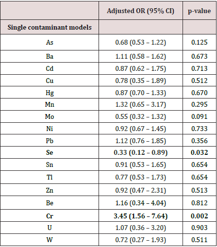
Table 4 and Figure 1 reveal and display that, Cr chromium at Cut off point >0.92 have AUC=0.747, statistical Sensitivity=80.0, statistical Specificity=60.8, PPV=13.6, NPV =97.5, Se selenium at cutoff point ≤35.4 have AUC=0.759, statistical Sensitivity=83.3, statistical Specificity=62.6, PPV=14.6, NPV =98.
Table 4: Relation between adverse neonatal outcome and fetal urinary production rate, adjusted for gestational age. FUPR: Fetal Urinary Production Rate; CSF: Cerebrospinal Fluid; NEC: Necrotizing Enterocolitis; IVH: Intraventricular Hemorrhage. •: Independent t-test.

Discussion
Trace metals and angiogenic biomarkers are of increasing research interest since investigative approaches in prior research efforts have revealed that angiogenic markers are reflecting the trophoblastic invasiveness process that is markedly impaired in preeclampsia pathological developmental course [19-21]. Furthermore it was observed that trace metals were observed to be of cornerstone importance to be maintained within normal safe acceptable physiological levels that would maintain the cellular metabolic and division activities that are well observed in the process of placentation and spiral artery remodeling ,on the other hand it was observed that the toxic impact of lead could impair the cellular apoptotic processes that are required for spiral artery remodeling besides it teratogenic impact. Preeclampsia although it is the disease of theories it is considered triggered by environmental exposure levels to various issues such as trace metals that interact in a manner that affect the oxidative stress process and early physiological development of the placenta [22,23].
An increased requirement for early screening and diagnosis of preeclampsia is crucial to upgrade the management protocols of this challenging obstetric clinical scenario [24,25]. A prior research group of investigators performed a research effort similar to the current study as regards the approach and methodology have revealed and displayed among their research results that there is raised risk for preeclampsia pathological development correlated to the detectability of chromium in the investigated urine early in gestation and a decreased clinical risk of preeclampsia development correlated to raised urinary Selenium denoting that selenium regarding its antioxidant properties is a safe guard against preeclampsia pathological development ,interestingly those research results show great harmony and similarity to the current study results denoting the clinical value of examining trace metals in gestation to predict clinical risk of preeclampsia [26,27]. Furthermore another research team of investigators gave observed that urinary cadmium, besides other toxic and essential trace metals, is correlated in a statically significant manner to reduced circulating PlGF levels and urinary copper is correlated to elevated sFlt-1 serum levels and an elevated sFlt-1/PlGF ratio, suggesting a linkage and correlation between physiologically impaired placentation process and clinical risk of preeclampsia those research observations have shown great harmony with the current study findings and could be justified by the fact that the placentation process is highly sensitive at cellular and molecular levels atha could be affected by any alterations in the environmental zone of placental development such as exposure to toxic metals [28,29]. Recent research efforts have displayed that chromium exposure causes raised expression of oxidative stress bio markers and apoptotic signaling within developing trophoblast tissues in experimental animals justifying the current study findings based on molecular-level research evidence the linkage between preeclampsia development and chromium toxic exposure levels [1,3,5,9].
Interestingly previous research efforts have observed that there is correlation between urinary cadmium and lead lower PlGF serum levels. Both Cd and Pb are trace metals found in contaminated air, soil, or water. Cd levels tend to be higher foods, e.g. rice and grains, and some drinking water systems denoting that the environmental pollution exposure is a cornerstone issue for preeclampsia clinical risk being raised within the affected population that relies on the geographical zone affected by the pollutant [2,10,13]. Another research meta-analyses have revealed that higher plasma or serum copper is correlated in a statically significant fashion to raise with preeclampsia risk ,those research findings could be justified by the fact that modest rise in copper levels could trigger the of reactive oxygen species raised productivity and oxidative stress, that are well demonstrated to be correlated to raised risk for preeclampsia development [15,19,26]. The current research study findings are well supported by prior molecular level observations that, toxic and essential metals have complex interactions within the placental tissue particularly in early phases of development. e.g. antagonistic molecular interactions between cadmium and selenium maternal serum levels affecting placental apoptotic gene expressive patterns [20,23,28].
Conclusion and Recommendations for Future Research
The current study provokes the clinical value for trace metal screening in detectability of preeclamptic risk of development ,however future research efforts are require to correlate the clinical risk of disease development according to the environmental trace metal levels ,besides future research efforts should be multicentric in fashion with larger sample sizes that could elucidate the best trace metals that could be implemented in screening to reveal the risk of developing preeclampsia.
References
- ACOG (2013) Hypertension in pregnancy. Report of the American College of Obstetricians and Gynecologists’ task force on hypertension in pregnancy. Obstet Gynecol 122(5): 1122-1131.
- Steegers EA, Von Dadelszen, Duvekot JJ, Pijnenborg R (2010) Pre-eclampsia. Lancet 376(9741): 631-644.
- Fisher SJ (2015) Why is placentation abnormal in preeclampsia? Am J Obstet Gynecol 213(4 Suppl): S115-122.
- M Timmermans S, Groot CJ, Russcher H, Lindemans J, Hofman A, et al. (2012) Angiogenic and fibrinolytic factors in blood during the first half of pregnancy and adverse pregnancy outcomes. Obstet Gynecol 119(6): 1190-1200.
- Mc Elrath TF, Lim KH, Pare E, Rich-Edwards J, Pucci D, et al. (2012) Longitudinal evaluation of predictive value for preeclampsia of circulating angiogenic factors through pregnancy. Am J Obstet Gynecol 207(5): 407.e1-7.
- Allen RE, Rogozinska E, Cleverly K, Aquilina J, Thangaratinam S (2014) Abnormal blood biomarkers in early pregnancy are associated with preeclampsia: a meta-analysis. Eur J Obstet Gynecol Reprod Biol 182: 194-201.
- Verlohren S, Herraiz I, Lapaire O, Schlembach D, Moertl M, et al. (2012) The sFlt-1/PlGF ratio in different types of hypertensive pregnancy disorders and its prognostic potential in preeclamptic patients. Am J Obstet Gynecol 206(1): 58.e1-8.
- Jameil NA (2014) Maternal serum lead levels and risk of preeclampsia in pregnant women: a cohort study in a maternity hospital, Riyadh, Saudi Arabia. Int J Clin Exp Pathol 7(6): 3182-3189.
- Wang F, Fan F, Wang L, Ye W, Zhang Q, et al. (2018) Maternal cadmium levels during pregnancy and the relationship with preeclampsia and fetal biometric parameters. Biol Trace Elem Res 186(2): 322-329.
- Brooks SA, Fry RC (2017) Cadmium inhibits placental trophoblast cell migration via miRNA regulation of the transforming growth factor beta (TGF-beta) pathway. Food Chem Toxicol 109(Pt 1): 721-726.
- Erboga M, Kanter M (2016) Effect of cadmium on trophoblast cell proliferation and apoptosis in different gestation periods of rat placenta. Biol Trace Elem Res 169(2): 285-293.
- Zhang X, Xu Z, Lin F, Wang F, Ye D, et al. (2016) Increased oxidative DNA damage in placenta contributes to cadmium-induced Preeclamptic conditions in rat. Biol Trace Elem Res 170(1): 119-127.
- Zhang Q, Huang Y, Zhang K, Huang Y, Yan Y, et al. (2016) Cadmium induced immune abnormality is a key pathogenic event in human and rat models of preeclampsia. Environ Pollut 218: 770-782.
- Ma Y, Shen X, Zhang D (2015) The relationship between Serum Zinc level and Preeclampsia: a meta-analysis. Nutrients 7(9): 7806-7820.
- Ferguson KK, McElrath TF, Meeker JD (2014) Environmental phthalate exposure and preterm birth. JAMA Pediatr 168(1): 61-67.
- Ferguson KK, McElrath TF, Cantonwine DE, Mukherjee B, Meeker JD (2015) Phthalate metabolites and bisphenol-A in association with circulating angiogenic biomarkers across pregnancy. Placenta 36(6): 699-703.
- Zwolak I, Zaporowska H (2012) Selenium interactions and toxicity: a review. Selenium interactions and toxicity. Cell Biol Toxicol 28(1): 31-46.
- Al-Saleh I, Al-Rouqi R, Obsum CA, Shinwari N, Mashhour A, et al. (2015) Interaction between cadmium (cd), selenium (se) and oxidative stress biomarkers in healthy mothers and its impact on birth anthropometric measures. Int J Hyg Environ Health 218(1): 66-90.
- Everson TM, Kappil M, Hao K, Jackson BP, Punshon T, et al. (2017) Maternal exposure to selenium and cadmium, fetal growth, and placental expression of steroidogenic and apoptotic genes. Environ Res 158: 233-244.
- Cantonwine DE, Meeker JD, Ferguson KK, Mukherjee B, Hauser R, et al. (2016) Urinary concentrations of bisphenol a and phthalate metabolites measured during pregnancy and risk of preeclampsia. Environ Health Perspect 124(10): 1651-1655.
- Maduray K, Moodley J, Soobramoney C, Moodley R, Naicker T (2017) Elemental analysis of serum and hair from pre-eclamptic South African women. J Trace Elem Med Biol 43: 180-186.
- Zhu Q, Zhang L, Chen X, Zhou J, Liu J, et al. (2016) Association between zinc level and the risk of preeclampsia: a meta-analysis. Arch Gynecol Obstet 293(2): 377-382.
- Xu M, Guo D, Gu H, Zhang L, Lv S (2016) Selenium and preeclampsia: a systematic review and meta-analysis. Biol Trace Elem Res 171(2): 283-292.
- Brooks SA, Martin E, Smeester L, Grace MR, Boggess K, et al. (2016) miRNAs as common regulators of the transforming growth factor (TGF)-beta pathway in the preeclamptic placenta and cadmium-treated trophoblasts: Links between the environment, the epigenome and preeclampsia. Food Chem Toxicol 98(Pt A): 50-57.
- Kim SS, Meeker JD, Carroll R, Zhao S, Mourgas MJ, et al. (2018) Urinary trace metals individually and in mixtures in association with preterm birth. Environ Int 121(Pt 1): 582-590.
- Cantonwine DE, Ferguson KK, Mukherjee B, Chen Y-H, Smith NA, et al. (2016) Utilizing longitudinal measures of fetal growth to create a standard method to assess the impacts of maternal disease and environmental exposure. PLoS One 11(1): e0146532.
- Ferguson KK, McElrath TF, Meeker JD (2014) Environmental phthalate exposure and preterm birth. JAMA Pediatr 168(1): 61-67.
- Laine JE, Ray P, Bodnar W, Cable PH, Boggess K, et al. (2015) Placental cadmium levels are associated with increased preeclampsia risk. PloS One 10(9): e0139341
- Elongi JP, Scheers H, Tandu-Umba B, Haufroid V, Buassa B, et al. (2016) Preeclampsia and toxic metals: a case-control study in Kinshasa, DR Congo. Environ health 15: 48.

Top Editors
-

Mark E Smith
Bio chemistry
University of Texas Medical Branch, USA -

Lawrence A Presley
Department of Criminal Justice
Liberty University, USA -

Thomas W Miller
Department of Psychiatry
University of Kentucky, USA -

Gjumrakch Aliev
Department of Medicine
Gally International Biomedical Research & Consulting LLC, USA -

Christopher Bryant
Department of Urbanisation and Agricultural
Montreal university, USA -

Robert William Frare
Oral & Maxillofacial Pathology
New York University, USA -

Rudolph Modesto Navari
Gastroenterology and Hepatology
University of Alabama, UK -

Andrew Hague
Department of Medicine
Universities of Bradford, UK -

George Gregory Buttigieg
Maltese College of Obstetrics and Gynaecology, Europe -

Chen-Hsiung Yeh
Oncology
Circulogene Theranostics, England -
.png)
Emilio Bucio-Carrillo
Radiation Chemistry
National University of Mexico, USA -
.jpg)
Casey J Grenier
Analytical Chemistry
Wentworth Institute of Technology, USA -
Hany Atalah
Minimally Invasive Surgery
Mercer University school of Medicine, USA -

Abu-Hussein Muhamad
Pediatric Dentistry
University of Athens , Greece

The annual scholar awards from Lupine Publishers honor a selected number Read More...


