Lupine Publishers Group
Lupine Publishers
Menu
ISSN: 2637-4544
Review ArticleOpen Access
Clinical Biomarkers and Biotechnology in Fertile Window Volume 2 - Issue 2
Murcia Lora José María*
- Functional Reproductive Medicine and Biotechnology, Independent Researcher Clinical Consulting G&E, Logroño, España, Spain
Received: July 04, 2018;; Published: July 10, 2018
Corresponding author: José María Murcia Lora, Functional Reproductive Medicine and Biotechnology, Independent Researcher Clinical Consulting G&E, Logroño, España, Spain
DOI: 10.32474/IGWHC.2018.02.000135
Abstract
The works destined to the recognition of the fertility in the fertile window have been carried out by means of different clinical studies, and medical devices that have been object of interest and of investigation at international level, in the last years. In this context, this article aims to expose some of the biophysical characteristics of cervical secretion that has influenced advances in biotechnology applied to the recognition of fertility. Cervical secretion is mainly reviewed as a biomarker applied to the recognition of fertility in the fertile window in this context.
Keywords: Fertile Window, Biotechnology, Fertility Awareness, Biomarkers, Ovulation, Menstrual Cycle, Cervical Secretion, Sterility, Subfertility, Infertility
Abbrevations: GNRH: Gonadotropin-Releasing Hormone, LH: Luteinizing Hormone, FSH: Follicle-Stimulating Hormone, SPD: Swiss Precision Diagnostics, ODE: Estimated Day for Ovulation
Introduction
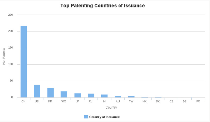
The recognition of fertility is increasingly important in prestigious scientific groups and in renowned international schools [1]. In this sense, we have tried to objectify the changes observed in the evolutionary process in the fertile window produced by the effect of estrogen and progesterone. Among the most relevant schools with this methodology are some authors such as; Hilgers[2], Stanfort[3], Scarpa [4], Aldecreutz[5], Ecochard[6] etc. These relevant contributions are based on the same clinical principle of fertile clinical window, which are based on the changes of cervical secretion pattern throughout the menstrual cycle. The fundamentals of this interest are justified in the perspectives that the application of the accumulated research of the basic principles of ovarian physiology pose. The pulsatile release of gonadotropinreleasing hormone (GNRH) stimulates the production of luteinizing hormone (LH) and follicle-stimulating hormone (FSH) in the anterior pituitary gland. These two hormones are responsible for stimulating the secretion of estrogen and progesterone at the level of the ovary, which justifies the physicochemical changes that occur in the cervical secretion [7]. Another aspect derived from clinical research in this field is the biotechnological implications derived from the biophysical properties of cervical secretion in the recognition of fertility. In this same line, the number of companies recognized by the scientific community in the development of devices related to the recognition of fertile time has increased. Among them: Swiss Precision Diagnostics (SPD) GmbH (Switzerland), Church & Dwight Co., Inc. (USA), Prestige Brands Holdings, Inc. (USA), Fairhaven Health, LLC (USA) ,HiLin Life of the product, Inc. (USA), Fertility Focus Limited (UK), and Geratherm Medical AG (Germany). According to a report recently published by Markets and Markets, the global market for devices, or so-called fertility tests, during the projected period of 2015 to 2020 is estimated to reach USD 151.0 million. Additionally, investment in this type of technology is forecast to grow at a compound annual rate of 7.5% during the 2015-2020 forecast period to reach USD 216.8 million in 2020 (Figure 1). Since 2015, to mention one of the recently studied intervals in time, the region between the United States and Canada had one of the largest shares in the global market of devices and tests for the recognition of fertility. The expectations in this respect are evident in the results obtained in devices developed to diagnose fertility tests in the mentioned regions, and it is expected that in Asia and the Pacific a greater annual growth will be reached (Figure 2). Another fact that stands out is the interest associated with the greater number of patent applications in devices for the recognition of fertility, with China leading the market, followed by the US office, as observed in the time interval included between 2007-2010 (Figure 3). Among the main applicants are Aronne Louis J with 3.8% of the applications, Kosasky; Harold J., President and Fellows of Harvard College, and The Regents of the University of California each with 1.4% of the applications.
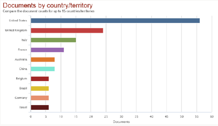
Figure 1: Scientific articles related to devices for the recognition of ferity. The United States has the largest number of publications, followed by England and Italy.

Figure 2: Patent applications by number in fertility devices. The majority were made in China, followed by the United States.

Biophysical Properties of Cervical Secretion and Biotechnology
Volume
One of the quantifiable observations of the cervical secretion, which characterizes its physical properties, has been the volume of it. It is known to increase near the peri-ovulatory period, observing the largest amount of secretion around day -1 and 0 in relation to the estimated day for ovulation (ODE), in natural cycles not subject to any type of treatment. This volume varies between the phases of the menstrual cycle [8].
Spinnbarkeit and Elasticity
The valuation in the changes in elasticity and filancia, have been recorded by means of descriptions that are carried out with protocols of different known authors, among them [2,4-6,9]. These schools associate descriptive properties with the trophic changes produced by estrogen and progesterone’s in cervical secretion. This fact, as the menstrual cycle progresses, allows to describe the variations that occur in the coloration, and elasticity, from an opaque and viscous sample, to a more transparent. The classic way of evaluating this parameter has been in general through the subjective evaluation in centimeters of the length of a quantity of secretion adhered to two separating surfaces [10]. The closest technology to assess this event has been classically the subjective determination by means of a graduated scale to the digital aperture in centimeters, in conjunction with the description of the observable properties [2,4,9].
Combination of Biophysical Parameters of Cervical Secretion: Mucoprotein Network and Hydration of the Cervical Mucus
The cervical secretion has been considered as a hydrogel composed of a liquid phase and a solid phase. The solid phase consists mainly of glycol proteins that are currently well characterized. The liquid phase is constituted by water and chemical and biochemical compounds such as salts, minerals, sugars, amino acids, lipids, protein chains, enzymes, etc. These components determine the main biophysical parameters of cervical secretion, which are mainly variations in quantity, appearance, transparency, viscoelasticity, and crystallization [11,12]. The aqueous component contains organic compounds such as glucose, amino acids, enzymes, soluble proteins, trace elements, and electrolytes including calcium, sodium, and potassium. However, the structural basis of cervical secretion is provided by mucins or large glycoproteins that intertwine to form meshes that behave differently with various biochemical and biophysical attributes. The main genes responsible for these properties, which determine the secretion of mucin are found in the endocervical epithelium and are MUC4, MUC5B, MUC5AC and MUC6 [13,14,15,16].
Biotechnology Applied to the Combination of the Mucoprotein Network and Rheological Properties of the Cervical Secretion
Within the flow term or rheological material subject to deformation, two properties are described, both viscosity and elasticity. These rheological properties of cervical mucus have been studied since the 1930s. The studies been done in terms of extensional rheology, in the sense of the study of flow and deformation caused by traction of a rheological material and the deformation resulting from a simple effort of secretion subject to cutting [16,17]. This methodology determines the observation in the middle of the cervical mucus cycle of translucent characteristics, less lumpy, more abundant and less viscous, than mucus in the early follicular phase, with a texture similar to that of egg white. Feature known as spinnbarkeit; or “spinning capacity” of a substance, or the capacity of a liquid that is stretched, which has been used as a test for the diagnosis of fertile time [11].The term spinnbarkeit has been coined to describe the elasticity during extension [16,18]. It has been possible to determine by electron microscopy accurately the changes in the filament of cervical secretion, which is maximum in the preovulatory phase, and can exceed 20cm in the ovulatory phase. The greater filancia in preovulatory phase, and quantification of its gradual ascent, has been objectified by valuation of the fibrils of glycol proteins, which behave as a mesh that retains a more or less liquid medium.At the beginning and at the end of the cycle, the diameter of the meshes does not exceed 0.2-0.5um in diameter Bar = 1um. During the follicular phase the pores of this network increase gradually under the effects of estrogen until reaching or exceeding 12um in diameter, Bar = 20um in the preovulatory period. This change and physical property that determines the fluidity and transparency of the cervical secretion can be quantifiable, due to the variability of the orifices regarding its relation with the fertile window, since its internal structure varies, which enlarges in porosity, allowing more or less fluidity [19]. In this context, one of the closest technologies that has been tried to determine this physical property, has probably been the methodology directed to the detection of hydration of the cervical mucus, in which the combination of fluidity, elasticity and hydration parameters is combined the cervical secretion. This biophysical property is apparently determined preferably by the concentration of MUC5B, which is highest during ovulation [16]. Regarding the analysis of this physical property, the probably closest biotechnological approach goes back to the studies of Kopito, who determines this physical property, by applying a detector that contains a light source, a photoreceptor and a light guide positioned to guide the light from the source to the photoreceptor. The light guide and the detector need to be inserted lightly into the external cervical os during use, for which an invasive procedure and its intracervical application is necessary. This technique involves internal manipulation, for the exploration of the sample. This type of invasive procedures makes it difficult to manipulate the sample, due to its intravaginal manipulation [20,21].
Commentary and Inventive Level in Biotechnology in Biophysical Properties of Cervical Secretion
One of the ways of assessing this qualitative leap to the biotechnological future is through the study of previously carried out antecedents as already mentioned. Another way of acting and being able to define a line of approach is to assess the capacity or inventive level in a network analysis of citations, as has been seen, which includes various analysis criteria to be considered in the valuation. Another valid criterion to corroborate the methodological success in the biotechnological search is the criterion of non-obviousness for a technician or professional moderately versed in the matter. This parameter evaluates the inventive capacity of a technique, by estimating the problem-solution binomial in the methodology used in the study of the antecedents of the state of the closest technique. The association of the determination of these parameters is based on the gradual evolution of the changes produced by the endocervical columnar epithelial cells in the cervical secretion, at the level of the endocervical canal before the production of estrogen and progesterone, which in the pre-ovulatory phase is composed up to 96% of water, and its quantification at the level of progressive physical change in combination of both fluidity, and elasticity quantified correlatively. These characteristics have not been achieved in any device, at the level of early stage of follicular phase, or any measure that determines the increase as it approaches the fertile window, where it increases the characteristics of transparency, quantity and filancia.
Probably the most classic system and method used for this purpose has been the method derived from the observation of the biophysical parameters of cervical secretion, which justifies the basis of many advances and developments based on the observation of the changes derived from estrogen. at the level of cervical secretion in the vulva and endocervical [2,4,9,12] which has been of interest and focus of attention in this article. However, since the classic pattern of recognition of Billings’ evolutionary pattern, its quantified characterization has not been possible [9]. Currently it is not possible through any device for commercial use, at low cost, or at a domestic level the possibility of determining the network that is formed, and the variations of the hormonal influx. It has also not been possible to assess the variability of fluidity in the lattice network of mucin. The valuation in the changes in elasticity and filancia of the cervical secretion has been registered through descriptions that are carried out with protocols widely known by renowned schools, as previously mentioned.The mentioned schools associate the descriptive properties as the menstrual cycle progresses, which allowdescribing variations in the coloration, from an opaque, more viscous sample, to become more transparent and more lined [2,6,8,9,12]. The classic way of evaluating this parameter has been in general by measuring in centimeters the length of a quantity of secretion adhered to two separating surfaces. The closest technology to assess this event has been classically the determination by means of a graduated scale to the digital aperture, in conjunction with the description of the observable properties. However, it is not possible to integrate a measurement and an objective assessment of the evolutionary changes of the biophysical variables of the cervical secretion.
Conclusion
The development of clinical methodologies and biotechnological tools in the study of the recognition of fertility is of global and current interest. It could be said that the binomial between the development of clinical methodologies and biotechnology in the study of the recognition of fertility has a promising future in the coming years in terms of the development of new technologies in the fertile window. The works destined to the recognition of the fertility by means of the valuation of clinical studies, and medical devices have been object of investigation to international level in the last years. It is expected that the technology developed in the diagnosis of ovulation, together with the advances in the methods applied to the recognition of fertility, will influence in the near future, to facilitate the conception among women with difficulties to become pregnant. Finally, the estimation of efforts in devices and methods as diagnostic tools in this field could reach criteria of novelty and inventive capacity, through analysis of patent citation networks, analysis of obviousness and state of the art, in techniques close to the technology that integrates the physicochemical variables of the cervical secretion. For this reason, the interest and development of biotechnological efforts that increase prospects in states of subfertility, or in situations associated with predicting receptive sperm capacity in the reproductive area, is justified.
References
- Stanford JB(2015) Revisiting the fertile window. FertilSteril 103 (5) : 1152-1153.
- Hilgers TW, Prebil AM(1979) The ovulation method-vulvar observations as an index of fertility. ObstetGynecol 53:12-22.
- Stanford JB, Smith KR, Dunson DB (2003) Vulvar mucus observations and the probability of pregnancy. ObstetGynecol101(6):1285-1293.
- Scarpa, Dunson DB, Colombo B(2006) Cervical mucus secretions on the day of intercourse: An accurate marker of highly fertile days. European Journal of Obstetrics & Gynecology and Reproductive Biology 125(1):72- 78.
- Adlercreutz H, Brown J, Collins W, Goebelsman U, Kellie A, et al. (1982) The measurement of urinary steroid glucuronides as indices of the fertile period in women. World Health Organization, Task Force on Methods for the Determination of the Fertile Period, special programme of research, development and research training in human reproduction. J Steroid Biochem17(6):695-702.
- Ecochard R, Duterque O, Leiva R, Bouchard T, Vigil P (2015) Selfidentification of the clinical fertile window and the ovulation period. FertilSteril. 103(5):1319-1325.
- Behre HM, Kuhlage J, Gassner C, Sonntag B, Schem C, et al. (2000) Prediction of ovulation by urinary hormone measurements with the home use ClearPlan Fertility Monitor: comparison with transvaginal ultrasound scans and serum hormone measurements. Hum Reprod15(12):2478-2482.
- Temprano H (2010) AplicacionesClínicas de la ventanafértilen los ciclosovulatorios. CURSO PRESYMPOSIUM. Bases y Aplicaciones de la fertilidadhumana. Indicadores de la fertilidad. DiagramaOdeblad. IX SimposioInternacional de Conciencia de Fertilidad Humana. La Coruña. España.
- Billings EL, Billings JJ, Brown JB, Burger HG (1972) Symptoms and hormonal changes accomplanying ovulation. Lancet 299(7745):282- 284.
- Elstein M (1982) Cervical mucus: its physiological role and clinical significance. Adv Exp Med Biol144:301-318.
- Clift AF (1945) Observations on Certain Rheological Properties of Human Cervical Secretion. Proc R Soc Med 39(1):1-9.
- Odeblad E (1997) Cervical mucus and their functions. J Irish coll Physicians and Curg26(21): 27-32.
- PapiM, ArcovioG, BompianiA, CastagnolaM, ParasassiT, et al. (2007) Globular structure of human ovulatory cervical mucus. FASEB J 21(14):3872-3876.
- Gipson IK, Spurr-Michaud S, Moccia R, Qian Zhan, Neil Toribara, et al. (1999) MUC4 and MUC5B transcripts are the prevalent mucin messenger ribonucleic acids of the human endocervix. Biology of Reproduction 60(1):58-64.
- Andersch-Björkman Y, Thomsson KA,Holmén Larsson JM, Ekerhovd E, Hansson GC(2007) Large scale identification of proteins, mucins, and their O-glycosylation in the endocervical mucus during the menstrual cycle. Mol Cell Proteomics 6(4):708-716.
- Gipson IK, Moccia R, Spurr-Michaud S, Argüeso P, Gargiulo AR, et al. (2001) The amount of MUC5B mucin in cervical mucus peaks at midcycle. Journal of Clinical Endocrinology and Metabolism 86(2):594- 600.
- McKinley GH(2005) Visco-elasto-capillary thinning and break-up of complex fluids. Polymer British Society of Rheology p. 1-48.
- Bansil R, Stanley E, La Mont JT (1995) Mucin biophysics. Annu Rev Physiol57:635-657.
- Chretien FC (1975) Preparation du mucus cervical e l’observation au microscope electronique e balayage. JMicrosc Biol Cell 24:23-44.
- Kopito LE, Kosasky HJ, Stugis SH, Lieberman BL, Shwachman H (1973) Water and electrolytes in human cervical mucus. Fertility and Sterility 24(7):499-506.
- Kopito LE, Kosasky HJ (1979) The Tackiness Rheometer determination of the Viscoelasticity of Cervical Mucus. Human Ovulation, ESF Hafez (Eds.), Elsevier/North-Holland: Amsterdam. 351-361.

Top Editors
-

Mark E Smith
Bio chemistry
University of Texas Medical Branch, USA -

Lawrence A Presley
Department of Criminal Justice
Liberty University, USA -

Thomas W Miller
Department of Psychiatry
University of Kentucky, USA -

Gjumrakch Aliev
Department of Medicine
Gally International Biomedical Research & Consulting LLC, USA -

Christopher Bryant
Department of Urbanisation and Agricultural
Montreal university, USA -

Robert William Frare
Oral & Maxillofacial Pathology
New York University, USA -

Rudolph Modesto Navari
Gastroenterology and Hepatology
University of Alabama, UK -

Andrew Hague
Department of Medicine
Universities of Bradford, UK -

George Gregory Buttigieg
Maltese College of Obstetrics and Gynaecology, Europe -

Chen-Hsiung Yeh
Oncology
Circulogene Theranostics, England -
.png)
Emilio Bucio-Carrillo
Radiation Chemistry
National University of Mexico, USA -
.jpg)
Casey J Grenier
Analytical Chemistry
Wentworth Institute of Technology, USA -
Hany Atalah
Minimally Invasive Surgery
Mercer University school of Medicine, USA -

Abu-Hussein Muhamad
Pediatric Dentistry
University of Athens , Greece

The annual scholar awards from Lupine Publishers honor a selected number Read More...


