Lupine Publishers Group
Lupine Publishers
Menu
ISSN: 2641-1652
Research Article(ISSN: 2641-1652) 
Visceral adiposity in Obese Diabetic Subjects: Influence of Age and Sex Volume 3 - Issue 4
Larbaoui K and Ghouini A*
- Laboratory of Physiology, Faculty of Medicine/CHU Blida, Algeria
Received: May 21, 2022; Published: June 02, 2022
*Corresponding author: Laboratory of Physiology-Faculty of Medicine/CHU Blida, Algeria
DOI: 10.32474/CTGH.2022.03.000170
Abstract
The distribution of body fat is actually more important than the total amount of fat mass when it comes to cardio-metabolic risk. Abdominal visceral fat is just one low share of fat mass, since the majority of fat is subcutaneous (80% in men, 90% in women), but seems to play a more important role than the rest of the adipose tissue. Indeed, the fatty tissue has 2 compartments with characteristics different metabolisms. In our work, we investigated the influence of age and sex on the propensity for the development of visceral fat in a target population such as obese diabetics. At the end of our study, it clearly appears that age is a risk factor for increased visceral adipose tissue and that men are more exposed than women, although the BMI is higher in the latter.
Obesity; Visceral Adiposity; BMI; Age; Sex; Cardio; Metabolic Risk
Introduction
Obesity is a public health problem. The prevalence of obesity is increasing in all industrialized countries. Android obesity is associated with excess cardiovascular mortality and type 2 diabetes, which justifies early management of this disease [1]. Visceral adipose tissue, via portal drainage, could be an important source of free fatty acids with complex metabolic effects: participation in hepatic lipogenesis, increase in hepatic neoglucogenic flux, decrease in metabolic clearance of insulin and participation in Peripheral insulin resistance via a Randle-type substrate competition phenomenon [2]. These phenomena increase the risk of cardiovascular diseases, cancers and major metabolic disorders [3]. To this end, we want to research the risk factors likely to favor the development of visceral adiposity and the unfavorable repercussions on the cardio-metabolic level.
Patients and Methods
Characteristics of the population
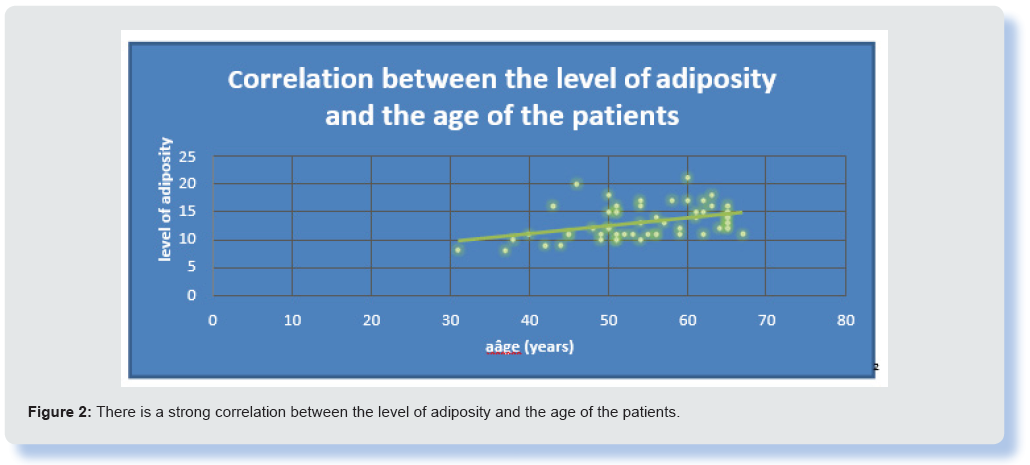
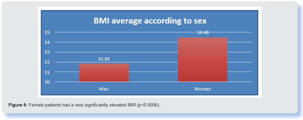
During this study we collected 55 obese diabetic adult patients of both sexes followed in consultation for metabolic diseases at the EPSP of Blida and the Physiology department of the N. Hamoud hospital of Hussein Dey grouped into three classes according to the level of visceral adiposity (Tables 1 & 2) (Figures 1 & 2). Anthropometric measurements for the calculation of body mass index (BMI) and impedance measurements to assess the level of visceral fat using the BF 508 type OMRON body composition monitor were undertaken (Table 3) (Figures 3 & 4). The variables associated with the level of visceral fat were researched by Statistical analysis was performed with SPSS software version 25 (IBM SPSS). The qualitative variable (sex) is described in numbers (percentage), the continuous variables (quantitative) are presented in the form of means +/- standard deviations. Student’s t test and anova test were used to compare quantitative variables. A p-value less than 0.05 is assumed to be statistically significant (Tables 4 & 5) (Figures 5 & 6).
Figure 1: More than 50% of the population has a high level of visceral adiposity and less than 10% has a normal level.
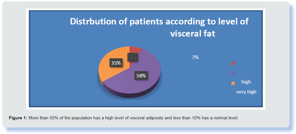
Figure 4: The patients with a very high level of visceral adiposity (15 -30) were very significantly older (p < 0.0001).
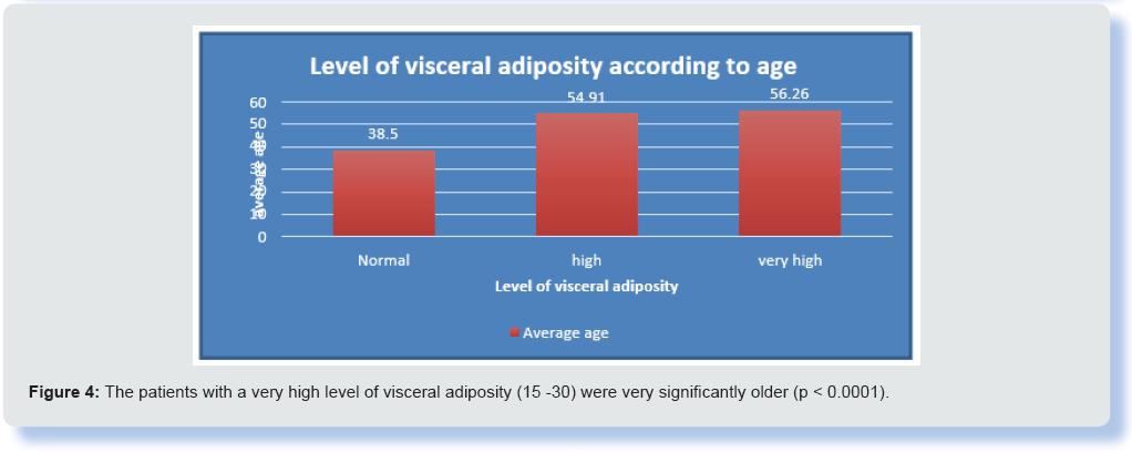
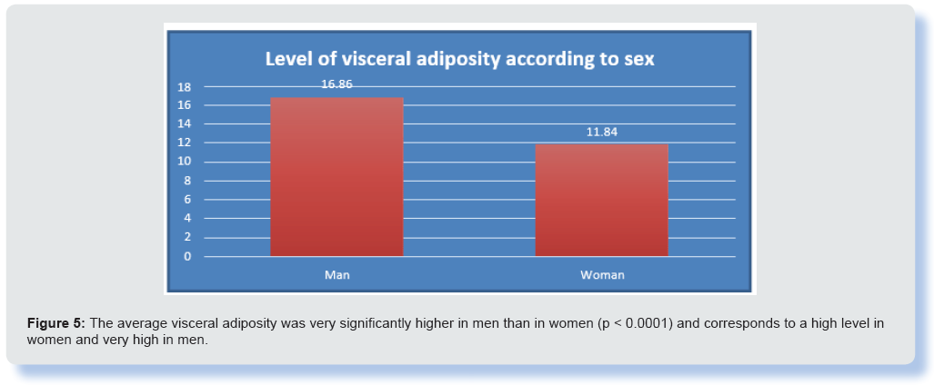
Figure 5: The average visceral adiposity was very significantly higher in men than in women (p < 0.0001) and corresponds to a high level in women and very high in men.
Results
Demographic data: There is a strong correlation between the level of visceral adiposity and the age of patients with an average age of 54.18 +/- 8.38 years.
Discussion
The importance of the regional distribution of adipose tissue as a factor closely associated with dyslipoproteinaemia and cardiovascular disease and type 2 diabetes has been highlighted by the scientific community interested in obesity and its complications [4]. Moreover, multivariate analyzes revealed that the amount of visceral adipose tissue measured by axial tomography was the variable most strongly correlated with different ratios of parameters used in the prediction of the risk of metabolic disorders [5,6]. Our results plead in favor of a relationship between age and the importance of visceral adiposity that may implicate sedentary lifestyle and the drop in the level of energy expenditure consistent with the current environment, which corroborates the data from the literature. However, the comparison of the BMI of the two sexes shows that there is no relationship between this index and the level of visceral fat due to the significant particularity of this visceral adipose tissue which may be present in subjects with an index of normal body weight and who may still be exposed to the metabolic and cardiovascular risks of obesity [7].
In contrast, there are a small number of android obese patients without increased visceral fat (1 in 3 cases) [8]. Moreover, contrary to generally accepted ideas, the subcutaneous adipose tissue also participates in the pool of easily mobilized free fatty acids in the obese [9]. Thus, the links between obesity and metabolic diseases could go through the facilitated availability of fatty acids from adipose tissue (visceral and subcutaneous) in otherwise genetically predisposed subjects [10].
Conclusion
This study clearly shows the role of age in the expansion of visceral adipose tissue; this trend is more marked in men than in women who generally have a higher BMI. Visceral fat exposes to more cardiometabolic risk than subcutaneous fat which, nevertheless, has a relative deleterious potential.
References
1 Ziegler O, Trebea A, Tourpe D, Böhme P, Quilliot D, et al. (2007) Tissu adipeux viscéral: rôle majeur dans le syndrome métabolique. Cah Nutr Diet 42(2): 85-89.
- Björntorp P (2000) Différence métabolique entre graisse viscérale et graisse abdominale sous- cutanée. Diabetes 26(3): 10.
- Direk K, Cecelja M, Astle W, Chowienczyk P, Spector TD, et (2013) The relationship between DXA-based and anthropometric measures of visceral fat and morbidity in women. BMC Cardiovasc Disord 13: 1-13.
- Kuk JL, Church TS, Blair SN, Ross R (2010) Measurement Site and the Association Between Visceral and Abdominal Subcutaneous Adipose Tissue with Metabolic Risk in Obesity 18(7): 1336‑1340.
- Katzmarzyk PT, Greenway FL, Heymsfield SB, Bouchard C (2013) Clinical Utility and Reproducibility of Visceral Adipose Tissue Measurements Derived from Dual-energy Xray Absorptiometry in White and African American Adults. Obes Silver Spring 21(11): 2221‑2224.
- Machann J, Thamer C, Schnoedt B, Haap M, Haring H-U, et al. (2005) Standardized assessment of whole-body adipose tissue topography by MRI. J Magn Reson Imaging JMRI 21(4): 455‑462.
- Sert C (1994) Obésité viscérale ou être obèse et de poids normal. Endocrinologie-Nutrition.
- Thomas EL, Saeed N, Hajnal JV, Brynes A, Goldstone AP, et (1998) Magnetic resonance imaging of total body fat. J Appl Physiol Bethesda 85(5): 1778‑1785.
- Smith SR, Lovejoy JC, Greenway F, Ryan D, deJonge L, et (2001) Contributions of total body fat, abdominal subcutaneous adipose tissue compartments, and visceral adipose tissue to the metabolic complications of obesity. Metabolism 50(4): 425‑435.
- Despres J P, Lesage M, Lemieux S, Prud’homme D (1995) Groupement de facteurs de risque pour les maladies cardio-vasculaires dans l’obésité viscé Implications thérapeutiques. Annales d’endocrinologie 56(2): 101-105.

Top Editors
-

Mark E Smith
Bio chemistry
University of Texas Medical Branch, USA -

Lawrence A Presley
Department of Criminal Justice
Liberty University, USA -

Thomas W Miller
Department of Psychiatry
University of Kentucky, USA -

Gjumrakch Aliev
Department of Medicine
Gally International Biomedical Research & Consulting LLC, USA -

Christopher Bryant
Department of Urbanisation and Agricultural
Montreal university, USA -

Robert William Frare
Oral & Maxillofacial Pathology
New York University, USA -

Rudolph Modesto Navari
Gastroenterology and Hepatology
University of Alabama, UK -

Andrew Hague
Department of Medicine
Universities of Bradford, UK -

George Gregory Buttigieg
Maltese College of Obstetrics and Gynaecology, Europe -

Chen-Hsiung Yeh
Oncology
Circulogene Theranostics, England -
.png)
Emilio Bucio-Carrillo
Radiation Chemistry
National University of Mexico, USA -
.jpg)
Casey J Grenier
Analytical Chemistry
Wentworth Institute of Technology, USA -
Hany Atalah
Minimally Invasive Surgery
Mercer University school of Medicine, USA -

Abu-Hussein Muhamad
Pediatric Dentistry
University of Athens , Greece

The annual scholar awards from Lupine Publishers honor a selected number Read More...


