Lupine Publishers Group
Lupine Publishers
Menu
ISSN: 2637-4749
Case Report(ISSN: 2637-4749) 
Echocardiography, Ultrasonography and Laboratory Findings of Left Ventricular Systolic Dysfunction and Right-Sided Congestive Heart Failure in A Neonatal Calf Volume 1 - Issue 5
Alper Erturk*, Murat Kaan Durgut, Amir Naseri and Mahmut Ok
- Selcuk University Faculty of Veterinary Medicine Department of Internal Medicine, Konya, Turkey
Received: August 20, 2018; Published: August 24, 2018
Corresponding author: Alper Erturk, Selcuk University Faculty of Veterinary Medicine Department of Internal Medicine, Konya, Turkey
DOI: 10.32474/CDVS.2018.01.000125
Abstract
A 14-days old female Holstein calve was referred to the Large Animals Hospital of the Faculty of Veterinary Medicine of Selcuk University with a history of inappetence, weight loss and lethargy. On the initial examination, severe anemic mucosal membranes bilateral distended jugular veins and 4/6 degree holo-systolic tricuspidal murmur were presented. Echocardiographic and ultrasonographic findings showed LV systolic dysfunction and Right sided congestive heart failure. The high levels of CK-MB and cardiac troponin I demonstrated severe cardiac injury.
Keywords: Heart failure; Neonatal Calf; Echocardiography; Cardiac Biomarkers
Introduction
Heart failure (HF) is the terminal event with cardiac disease and occurs when initial cardiac and neuro-hormonal compensatory mechanisms are overwhelmed [1]. Clinical signs of HF in cattle include syncope, exercise intolerance and weakness [2]. Congestive heart failure (CHF) can be distinguished from HF by the presence of congestion, effusion and edema, which are the result of increased hydrostatic pressure and fluid retention [3]. The prognosis is traditionally said to be guarded to poor in bovine HD cases [4,5] but is slightly better in those cases of HD without clinical signs of HF [6]. Left ventricular systolic dysfunction commonly determined by reduction of ejection fraction (EF), fractional shortening (FS) or stroke volume (SV) [7]. There is many literature information about left ventricular systolic dysfunction in human medicine in varies of diseases [8, 9]. But there is little information about LV systolic dysfunction in neonatal calves [10]. Right-sided congestive heart failure (RHF), also known as high altitude disease or brisket disease, is initiated by hypoxia-induced pulmonary arteriolar narrowing.
Vessel narrowing increases resistance to blood flow, mean pulmonary arterial pressure and ultimately the risk of RHF [11,12]. Other causes such as pericardial effusion, tricuspid endocarditis or lymphoma of the right atrium can lead to RHF [3]. Diagnosis of HF in cattle is based on clinical signs, cardiac auscultation, electrocardiography, echocardiography, thorax radiography and laboratory findings [13]. It is of great interest to make a precise diagnosis in the field, as it permits rapid evaluation and decision making regarding treatment options and avoids wasteful supportive treatments in the case of a low-value animal. Ultrasonography is a good choice for imaging and describing most heart diseases [14]. Furthermore, cardiac ultrasound examination can be performed easily in field conditions with high sensitivity and specificity [15]. Previously, it has been used successfully in the field diagnosis [16, 17].
Case History and Clinical Observations
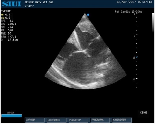
A 14-days old female Holstein calve was referred to the Large Animals Hospital of the Faculty of Veterinary Medicine of Selcuk University with a history of inappetence, weight loss and lethargy. On the initial examination, the calf was in sternal position with mild dehydration (6percent), severe anemic mucosal membranes and prolonged capillary refill time (7sec). Examination of cardiovascular system presented bilateral distended jugular veins, tachycardia (140bpm), tachypnea (80bpm) and 4/6 degree holosystolic tricuspidal murmur. Transthoracic echocardiography using of right parasternal long and short-axis, and left apical windows was performed to evaluate left and right ventricles. The echocardiographic findings included a hypokinetic LV with decreased ejection fraction (EF) (40 per cent), stroke volume (SV) (8ml) and cardiac index (CI) (910ml/kg/m2). Also, there was a dilated RV (RV:LV ratio> 0,6) and RA. Systolic pulmonary arterial pressure estimated by tricuspid insufficiency (TRVmax 2+RAP) and showed a moderate PAH (46mmHg) [18,19]. There was no pulmonary valve stenosis or left to right intracardiac shunts (Figures 1-3).
Figure 1: M-mode right parasternal short axis view of LV shows severe reduction of LV systolic function (EF: % 40).
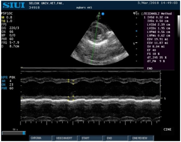
Figure 3: Liver ultrasonography shows congestion, increased echogenicity of its parenchyma and the dilatation caudal vena cava (CVC).
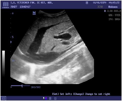
The ultrasonographic examination was performed using a 3.5-5MHz convex transducer probe. Ultrasonographic findings included hepatomegaly and congestion with increased echogenicity of its parenchyma and the caudal vena cava (CVC) dilated and did not collapsed during inspiration. The portal and hepatic veins (HV) were also dilated. These abnormalities have been attributed to cardiac insufficiency. Blood sampling performed from vena jugularis for blood gas analysis, CBC and serum biochemistry. Laboratory findings showed state of septicemia, severe anemia, severe pre-renal azotemia, metabolic acidosis and significant increases of cardiac biomarkers.
Treatment
In the treatment of fluid and respiratory acidosis, 0.9% NaCl solution was administered intravenously with sodium bicarbonate NaHCO3 solution. In addition, intravenously treated with Marbofloxacin 5mg/kg BW (Marbocyl 10%, Novakim), Meloxicam 0.5mg/kg BW (Maxicam, Sanovel), Furosemide 2mg/ kg BW, twice daily (Diuril, Vetas). Medications containing iron and B12 (Fercobsang, Novakim) were administered for the treatment of anemia in the patient. Oxygen therapy was applied for supportive treatment. Despite treatment, the patient died after 12 days.
Discussion
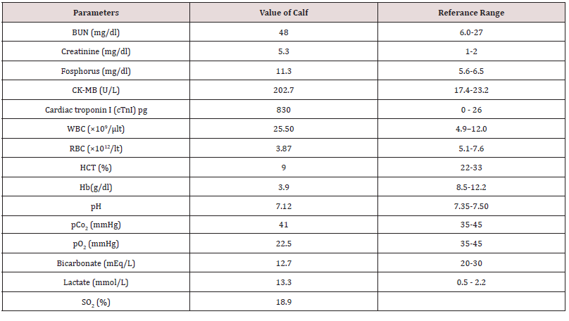
Heart failure (HF) is the terminal event of heart disease and is the consequence of progressive, adaptive changes in the neurohormonal axis that have multiple negative effects on the myocardium and cardiac output [2]. Diagnosis of heart failure is based on echocardiographic, ultrasonographic and laboratory findings. In this case echocardiographic findings of left ventricle showed a severe reduction of contractility ability of the left ventricle (EF). Previous studies in human medicine and veterinary medicine suggested that inflammatory state and septicemia can lead to left ventricular systolic dysfunction [8,10]. Our laboratory data showed that there is a state of septicemia in the calf and it may be the main causes of LV systolic dysfunction. Right-sided heart failure is one of the complications of pulmonary hypertension, pericarditis, myocarditis and tricuspid insufficiency in cattle [13]. In this case, there were no signs of pericarditis or primary tricuspid insufficiency but RV:LV ratio and Doppler findings of tricuspid valve regurgitation showed that there was a moderate PHT. In our opinion, because of any visible tricuspid and pulmonary valve abnormality, induction of PVR due to unknown etiological agent (eg. pulmonary embolism, idiopathic pulmonary hypertension) could leads to increases right ventricle intra-cardiac pressure and severe dilatation of RV, RA and so, right-sided congestive heart failure (Table1).
One of the other methods for confirm the right heart failure is liver ultrasonography. In the right heart failure because of increases in right atrial pressure, increase of pressure in the CVC is inevitable and it can see as dilated CVC and HV [20]. Our echocardiography and ultrasonography findings showed that because of severe volume over-load of right ventricle and right atrium, there was a severe induction of CVC pressure and dilatation of hepatic veins and probably hepatic congestion. Cardiac troponins and cardiac specific isoenzyme of CPK and CK-MB are the most useful of cardiac biomarkers for detecting myocardial injury in the calves [21-23]. Our data shows that there is a drastic elevation in the levels of cardiac troponin I and CK-MB. In our opinion, because of state of septicemia and right ventricular overload, it makes the myocardial damage inevitable and interpretation with echocardiographic finding together confirmed the severe circulatory failure and death in this case. In conclusion, left ventricular systolic dysfunction and right-sided heart failure in neonatal calves can raises from different etiological agents. Echocardiography, ultrasonography and laboratory findings can help the practitioners to get new perspectives from diagnosis to the prognosis and treatment in neonatal calves with heart failure.
References
- Colucci WS, Braunwald EB (2005) Pathophysiology of heart failure (7nd edn.), Braunwald’s Heart Disease: A Textbook of Cardiovascular Medicine, Elsevier Saunders, USA, pp. 509-538.
- De Morais HA, Schwartz DS (2005) Pathophysiology of heart failure (6nd edn.), Textbook of Veterinary Internal Medicine, Elsevier Saunders, USA, pp. 914-940.
- Reef VB, McGuirk SM (2002) Diseases of the cardiovascular system (3nd edn.). Large Animal Internal Medicine, Mosby, USA, pp. 443-478.
- Nart P, Thompson H, Barrett DC, Armstrong SC, McPhaden AR (2004) Clinical and pathological features of dilated cardiomyopathy in Holstein– Friesian cattle. Veterinary Record 155(12): 355-361.
- Buczinski S, Fecteau G, DiFruscia R (2006) Ventricular septal defects in cattle: 25cases. Canadian Veterinary Journal 47(3): 246-252.
- Buczinski S, Francoz D, Fecteau G (2006b) Congestive heart failure in cattle: 59cases (1990–2005). In: World Buiatrics Congress, Nice France OS18-1.
- Chengode S (2016) Left ventricular global systolic function assessment by echocardiography. Annals of cardiac anaesthesia 19(Suppl 1): S26.
- Vieillard-Baron A, Caille V, Charron C (2008) Actual incidence of global left ventricular hypokinesia in adult septic shock. Crit Care Med 36(6): 1701-1706.
- Cleland JGF, Torabi A, Khan NK (2005) Epidemiology and management of heart failure and left ventricular systolic dysfunction in the aftermath of a myocardial infarction. Heart 91(2): 7-13.
- Naseri A, Turgut K, Sen I, Ider M, Akar A (2018) Myocardial depression in a calf with septic shock. Veterinary Record Case Reports 6: e000513.
- Alexander AF, Jensen R (1963a) Pulmonary vascular pathology of high altitude induced pulmonary hypertension in cattle. Am J Vet Res 24: 1112-1122.
- Alexander AF, Jensen R (1963b) Pulmonary arteriographic studies of bovine high mountain disease. Am J Vet Res 24: 1094-1097.
- Buczinski S, Rezakhani A, Boerboom D (2010) Heart disease in cattle: diagnosis, therapeutic approaches and prognosis. The Veterinary Journal 184(3): 258-263.
- Buczinski S (2009) Cardiovascular ultrasonography in cattle. Vet ClinNorth Am Large Anim Pract 25(3): 611-632.
- Braun U (2009) Ultrasonography of the gastrointestinal tract in cattle. Vet Clin North Am Large Anim Pract 25(3): 567-590.
- Mohamed T, Buczinski S (2011) Clinicopathological findings and echocardiographic predication of the localisation of bovineendocarditis. Vet Rec 169(7): 180.
- Mohamed T (2010) Clinicopathological and ultrasonographicfindings in 40 water buffaloes (Bubalus bubalis) with traumaticpericarditis. Vet Rec 167(21): 819-824.
- Masuyama T, Kodama K, Kitabatake A, Sato H, Nanto S, et al. (1986) Continuous-wave Doppler echocardiographic detection of pulmonary regurgitation and its application to noninvasive estimation of pulmonary artery pressure. Circulation 74: 484-492
- Bossone E, Dandrea A, Dalto M, Citro R, Argiento P, et al. (2013) Echocardiography in pulmonary arterial hypertension: from diagnosis to prognosis. Journal of the American Society of Echocardiography 26(1): 1-14.
- Turgut K (2017) Klinik kedi ve köpek kardiyolojisi. (1st edn.) Nobel Tip Kitabevleri, Turkey.
- Peek SF, Apple FS, Murakami MA, Crump PM, Semrad SD (2008) Cardiac isoenzymes in healthy Holstein calves and calves with experimentally induced endotoxemia. Can J Vet Res 72(4): 356-361.
- Karapinar T, Dabak DO, Kuloglu T, Bulut H (2010) High cardiactroponin I plasma concentration in a calf with myocarditis. Can Vet J 51: 397-399.
- Aydogdu U, Yıldız R, Guzelbektes H, Coskun A, Sen I (2017) Cardiac biomarkers in premature calves with respiratory distress syndrome. Acta veterinaria Hungarica 64(1): 38-46.

Top Editors
-

Mark E Smith
Bio chemistry
University of Texas Medical Branch, USA -

Lawrence A Presley
Department of Criminal Justice
Liberty University, USA -

Thomas W Miller
Department of Psychiatry
University of Kentucky, USA -

Gjumrakch Aliev
Department of Medicine
Gally International Biomedical Research & Consulting LLC, USA -

Christopher Bryant
Department of Urbanisation and Agricultural
Montreal university, USA -

Robert William Frare
Oral & Maxillofacial Pathology
New York University, USA -

Rudolph Modesto Navari
Gastroenterology and Hepatology
University of Alabama, UK -

Andrew Hague
Department of Medicine
Universities of Bradford, UK -

George Gregory Buttigieg
Maltese College of Obstetrics and Gynaecology, Europe -

Chen-Hsiung Yeh
Oncology
Circulogene Theranostics, England -
.png)
Emilio Bucio-Carrillo
Radiation Chemistry
National University of Mexico, USA -
.jpg)
Casey J Grenier
Analytical Chemistry
Wentworth Institute of Technology, USA -
Hany Atalah
Minimally Invasive Surgery
Mercer University school of Medicine, USA -

Abu-Hussein Muhamad
Pediatric Dentistry
University of Athens , Greece

The annual scholar awards from Lupine Publishers honor a selected number Read More...


