Lupine Publishers Group
Lupine Publishers
Menu
ISSN: 2637-4749
Research Article(ISSN: 2637-4749) 
Cross Sectional Study of Fibromatous Epulis in A Buffalo Herd Volume 1 - Issue 5
S Krishna kumar*1 ,R Madheswaran2,M Ranjith kumar3,B Poovarajan4 ,S Kavitha3and Selvaraj1
- 1Department of Veterinary Medicine, Veterinary College and Research Institute, India
- 2Department of Veterinary Pathology, Veterinary College and Research Institute, India
- 3Department of Veterinary clinical medicine, Madras Veterinary College, India
- 4Department of Veterinary Microbiology, Veterinary College and Research Institute, India
Received: July 26, 2018; Published: August 06, 2018
Corresponding author: S Krishna Kumar, Department of Veterinary Medicine, Veterinary College and Research Institute, India
DOI: 10.32474/CDVS.2018.01.000123
Abstract
Fibromatous epulis, is a rare growth affecting the gingival mucosa of neonates. It is benign condition seen more frequently in females with multiple Epuli occurring in only 10% of cases. The cause and origin of fibromatous epulis remains unclear. This clinical investigations discussed about the epidemiology, clinical outcome, concurrent infection, biochemical and complete blood profile of a buffalo calf positive for epulis. Epulis arising from the upper and lower gingival margin, which were successfully managed therapeutically.
Keywords: Fibromatous Epulis; Buffalo Calf; Anemia; Toxacara
Introduction
Gingival masses of all types, many of which are of tooth germ origin, are common in dogs, but occur less frequently in other species [1]. The term epulis is used inconsistently for localized exophytic gingival growths, both reactive and neoplastic [2] and has been applied to various lesions including fibromatous epulis of periodontal ligament origin, acanthomatous ameloblastoma and peripheral giant cell granuloma [3]. The gingival tumours interfere with normal mastication of feed because of their location and pain sensation induced in adjoining areas and because of this reason animals undergo anorectic condition. This research paper reporting the epidemiology, concurrent infection, laboratory profile and clinical outcome of epulis in an organized buffalo farm.
Materials and Methods
Location
This research work was carried out in a district livestock farm, Thanjavur have 86 numbers of adult graded buffaloes and their calves during 2015 and 2016. Organized buffalo farm with good health care packages includes regular deworming and vaccination for Foot and Mouth Disease periodically.
Epidemiologic Investigations
During disease investigation farm level during March of 2015 and 2016, animals were examined and clinical samples with epidemiologic history were collected.
Samples Collection
Clinical samples like whole blood, serum, dung samples were subjected to complete blood analysis, biochemical profile, helminthes and microbial load were assessed. The mass was fixed in 10% neutral buffered formalin solution, embedded in paraffin sectioned at five-micrometer thickness and stained with routine hematoxylin and eosin (H&E).
Results
Animals in the flock with age from one month to 13 months includes both male and female. On clinical examination, most of the animals showed under weight for their respective age, condition of the animal is fair, mucous membrane showed congestion, Skin and coat is rough and dry (Scaly). Out of 26 buffalo calves born, 10 showed enteritis with yellowish colour with severe dehydration. Out of 10 animals six animals were recovered and two animals not responded to routine medical management and they were died. Out of ten animal three calves were showed growth in the gum region and samples collected for histopathological examination revealed that this was a case of fibromatous epulis. As per the record, average birth weight in 2014 is 22kgs in male calf and 25kgs in female calves. But in the year 2015 average birth weight in male calf is 28kgs and female calf is 27kgs. Growth rate in buffaloes seems to be 360gms /day. Complete blood count values showed anaemia and thrombocytosis and blood smear examination revealed no blood parasites. Biochemical analysis showed hyperproteinemia with elevated globunemia. Dung Examination revealed eggs of Amphistomes, Toxocara spp and Eimeria spp. In electrolyte analysis total calcium level was lower in animals affected with fbiromatous epulis. Microbiological examination of dung samples revealed E.Coli, Bacillus and Pseudomonas spp (Tables 1-4).
Discussion
Gingival masses of all types, many of which are of tooth gum origin, are common in dogs, but occur less frequently in other species [1]. The term ‘epulis’ from the Greek words ‘epi’ –over– and ‘oulon’ –gums was first used by Virchoff in 1864. The term epulis is used inconsistently for localized exophytic gingival growths, both reactive and neoplastic [2]. The affected calf belongs to an organized buffalo farm with good record keeping. The clinical history were poor weight gain and birth weight, in appentence and dehydration were because of the gingival tumors interfere with normal mastication of feed. Anemia and thromboctosis were noticed because of presence of Toxacara spp and Eimeria spp which causes blood loss periodically. The complete blood count showed positive for anemia which includes lower hemoglobulin level and RBC count. Moreover, the differential count also showed higher percentage of esinophil count which again proved the ongoing helminthic infection in the system. Higher total protein level was mainly because of elevated globulin level which in turn witnessed for chronic nature of the disease.
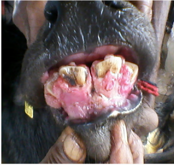
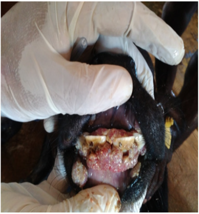
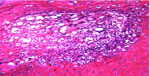
Microscopic examination of a gingival growth was diagnosed as fibromatous epulis. The localization, gross and histopathological findings of the present case are in agreement with other reports in dogs, cats and captive lion [3-5]. Histopathologically the revealed eosinophilic osteoid tissue surrounded by fibrous stroma interspaced with a few blood vessels (Figure 1). The dense collagen fiber had a high cellularity composed of small stellate to spindle shaped fibroblasts (Figure 2). The engorged, necrosis and mononuclear cells infiltration were observed (Figure 3). Nucleus revealed pleomorphism and increased nuclear: cytoplasmic ratio. Younger age groups like less than three months were mostly affected than adult and he also observed that because of their location and pain sensation induced in adjoining areas [1] and at times animal goes anorectic also noticed in this research (Figure 4). Hyperglobunemia also indicates the chronic nature of the inflammatory reaction in fibromatous epulis which was supported by histopathology of the gingival growth revealed the eosinophilic osteoid tissue surrounded by fibrous stroma and high cellulaity composed of small stellate to spindle shaped fibroblasts. Lower electrolytes value particularly calcium seems because of lower feed intake and poor mastication process by the mass interference in the mouth. The mass in the mouth which attracts higher microbial load in the host which also worsen the condition with diarrhea in the fibromatous epulis cases (Figure 5)
Figure 1: Histopathology of the gingival growth revealed the eosinophilic osteoid tissue surrounded by fibrous stroma. H&E. 100x.
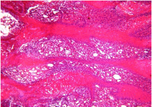
Figure 2: Histopathology of the gingival growth revealed high cellularity composed of small stellate to spindle shaped fibroblasts. H&E. 400x.
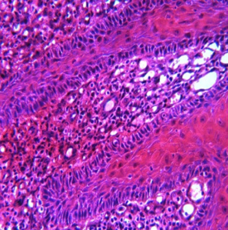
Figure 3: Histopathology of the gingival growth revealed necrosis and mononuclear cells infiltration. H&E. 400x..
Acknowledgement
The authors duly acknowledge the Dean, Veterinary college and research Institute, Orathanandu, Tamil nadu, India.
References
- Jubb KVF, Kennedy PC (1970) Pathology of Domestic Animals (2nd edn.) London, Academic Press.
- Gardner DG (1996) Epulides in the dog: a review. J Oral Pathol Med 25(1): 32-37.
- De Bruijn ND, Kirpensteijn J, Neyens IJS, Van Den Brand, JMA, Van Den Ingh (2007) A clinicopathological study of 52 feline epulides. Vet Pathol 44(2): 161-169.
- Head KW, RW Else, RR Dubielzing (2002) Tumors and tumor like lesions of vascular tissue: Tumors of Domestic Animals (4th edn.) Iowa State Press.
- Castro MB, Barbeitas MM, Borges TJ, Bonorino RP, Ramos RR, et al. (2011) Fibromatous epulis in a captive lion (Panthera leo). Brazilian J Vet Pathol 4(2): 150-152.

Top Editors
-

Mark E Smith
Bio chemistry
University of Texas Medical Branch, USA -

Lawrence A Presley
Department of Criminal Justice
Liberty University, USA -

Thomas W Miller
Department of Psychiatry
University of Kentucky, USA -

Gjumrakch Aliev
Department of Medicine
Gally International Biomedical Research & Consulting LLC, USA -

Christopher Bryant
Department of Urbanisation and Agricultural
Montreal university, USA -

Robert William Frare
Oral & Maxillofacial Pathology
New York University, USA -

Rudolph Modesto Navari
Gastroenterology and Hepatology
University of Alabama, UK -

Andrew Hague
Department of Medicine
Universities of Bradford, UK -

George Gregory Buttigieg
Maltese College of Obstetrics and Gynaecology, Europe -

Chen-Hsiung Yeh
Oncology
Circulogene Theranostics, England -
.png)
Emilio Bucio-Carrillo
Radiation Chemistry
National University of Mexico, USA -
.jpg)
Casey J Grenier
Analytical Chemistry
Wentworth Institute of Technology, USA -
Hany Atalah
Minimally Invasive Surgery
Mercer University school of Medicine, USA -

Abu-Hussein Muhamad
Pediatric Dentistry
University of Athens , Greece

The annual scholar awards from Lupine Publishers honor a selected number Read More...


