Lupine Publishers Group
Lupine Publishers
Menu
ISSN: 2637-4749
Research Article(ISSN: 2637-4749) 
Characteristic Cellular of the Vaginal Epithelium of Llamas (Lama glama) Captive in the State of Jalisco Volume 3 - Issue 2
De La Cruz Baltazar Eliab*
- Department of Veterinary Sciences, Zoológico Rancho Bonito, Mexico
Received: November 15, 2019; Published: December 03, 2019
Corresponding author: De La Cruz Baltazar Eliab, Department of Veterinary Sciences, Zoológico Rancho Bonito, Mexico
DOI: 10.32474/CDVS.2019.03.000157
Abstract
With the objective of contributing data regarding the characteristic cellular forms in vaginal smear of Llamas (Lama glama) and to determine their structural forms in different physiologic states, pregnant, nurslings and you empty, they were carried out vaginal smear of 15 Llamas in captivity condition in the State of Jalisco, Mexico. In the empty Llamas of the 10 to the 30 days post weaning the superficial cells spread to go down from 41% to 7% of presence and the 30 days post weaning the intermediate cells they spread to ascend from 58% to 98% of presence. In the pregnant Llamas in the first month of gestation the naviculars cells were observed in more percentage 86% and the cells flakes in smaller presence 10%, while to the 9° month of gestation increase the presence of cells flakes of 10% to 44% and they diminished the naviculars of 86% to 55%. In the Llamas in nursing of the 10 to the 30 days of nursing the cells flakes spread to ascend from 16% to 54%, the opposite happened to the intermediate cells that spread to go down from 83% to 42% to the 30 days of nursing. The cellular differences in the vaginal epithelium of the Llamas could serve like a tool in the diagnostic of gestation and fertility.
Keywords:Camelids; vaginal cytology; gestation; nursing
Introduction
The principle of the vaginal cytology is based on identifying the type and the percentage of cells in the different stages of the cycle estral, since the hormonal changes of the vagina during the cycle are reflected in the morphology of their cells epitelials[1]. The vaginal cytology is used in most of the species to determine stages of the cycle estral in which is the female, for what is a procedure broadly used in veterinary medicine, [2], and it is used with a lot of effectiveness to determine stadiums of the cycle estral in dogs and cats [3]Stornelli[4] mentions that in the canines with estro 80% of superficial cells is observed, while in the cats with estro 88% of superficial cells is observed [5], while in the necklace Pecarí(Tayassutajacu). Mayor et al.[6] found 60% of superficial cells in estro in the vaginal smear of Alpacas (Vicugnapacos). In a work it found differences in the vaginal epithelium among empty and pregnant Alpacas observing similar cells to other species[7]. The camelids has an unique ovarian cycle, they are induced ovuladores, but their hormonal physiology differs of other ovuladores induced as the rabbit or the cat, [8,9] and they don't present estacionality for photoperiod[10]. The camelids are considered as seasonal reproducers in its natural habitat during the warmest, humid months and of more forage readiness[11]. The vagina of the Llamas measures from 15 to 25cm from the hymen to the cervix and approximately 5 diameter cm [12], these characteristics are necessary to understand its ignorance since it takes us to have a poor yield of breeding, compared with other conventional species[13].
For such a reason the present study intends to carry out a classification at cellular level in the characteristic ways in vaginal smear of Llamas (Lama glama) captive, and to determine its structural forms in different physiologic states as a tool of diagnose reproductive.
Materials and Methods
15 females were chosen under captivity conditions belonging to hatcheries peculiar of the state of Jalisco, Mexico, with a corporal condition, weigh and age average of 4-5, 115±8 kg and 4.5±1.5, of which 9 were pregnant, 3 empty and 3 nurslings. For the taking of the samples we use the technique described by Wave [14] where we use a hyssop of soaked sterile cotton with solution of chloride of sodium to 0.9% which 8 cm was introduced inside the vagina carrying out rotational movements against the vaginal wall with the purpose of extracting cells that come off in a natural way rotating the hyssop on a portaobjets, later on noticing with ethylic alcohol of 90°, for the tint that of Giemsa it was preferred and you proceeded that recommended by Lynch [15] where once fixed with alcohol it allows to dry off and it dives for a while in the coloring Giemsa from 15 to 30 minutes, later that washes himself with distilled water and dried off to the environment. The observation to the microscope one carries out with increase of 100X and immersion oil, the opposing cells were classified following the approach of Schutte, [16] in you scale, intermediate, naviculars and superficial.
Results
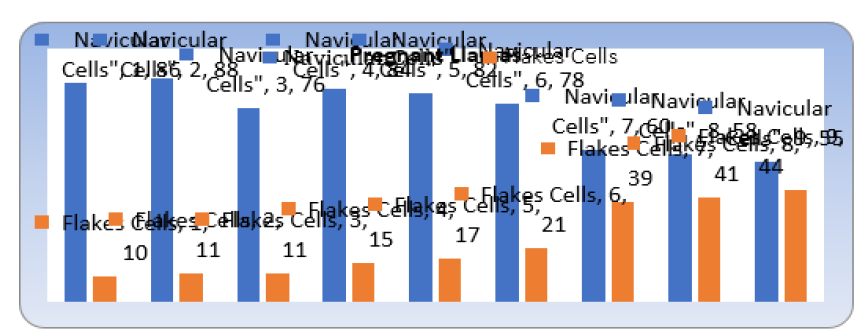
Figure 1: The following figure shows us the cellular activity from the first month of gestation up to the ninth in the 9 copies.

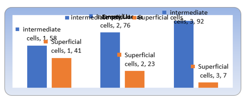
Figure 2: That shows us the cellular differences in the 3 copies of Llamas revised to the 10, 20 and 30 days search it weans.

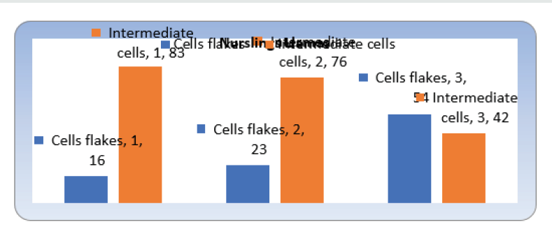
As it is appreciated in the Figures 1,2&3 previous to the first month of gestation the naviculars cells were observed in bigger percentage 86% and the flakes cells in smaller presence 10%, the opposite to the 9° month of gestation where it increase the presence of flakes cells of 10% to 44% and they diminished the naviculars of 86% to 55%. One can observe that of the 10 to the 30 days post weaning the superficial cells spread to go down from 41% to 7% of presence and the 30 days post weaning the Intermediate ones they spread to ascend from 58% to 98%, what resembles each other with the cat tames that it not indicates the existence of levels estrogenicos in the one post weaning early, [17]. As it is observed in the previous graph, of the 10 to the 30 days of nursing the cells flakes spread to ascend from 16% to 54% and the opposite happened to the intermediate cells that spread to go down from 83% to 42% to the 30 days.
Figure 3: Where show us the cellular characteristics in the 3 Llamas in nursing, from the 10 days up to the 30.

Discussion and Conclusion
In the pregnant Llamas in the first month of gestation the naviculars cells were observed in bigger percentage 86% and the flakes cells in smaller presence 10%, while to the 9° month of gestation increase the presence of flakes cells of 10% to 44% and they diminished the naviculars of 86% to 55%. In the empty Llamas of the 10 to the 30 days post weaning the superficial cells spread to go down from 41% to 7% of presence. To the 30 days post weaning in the empty Llamas the Cells intermediate spread to ascend from 58% to 98% of presence. as long as in the Llamas in nursing of the 10 to the 30 days of nursing the flakes cells spread to ascend from 16% to 54%. The opposite happened to the intermediate cells that spread to go down from 83% to 42% to the 30 days of nursing. The cellular differences in the vaginal epithelium of the Llamas could serve like a tool in the diagnostic of gestation and fertility.
Conflict of Interest
There are not conflict of interest exists.
References
- Arcila V, Serrano Novoa C, Hernández M (2005) Estandarización de la citología vaginal exfoliativa correlacionando los niveles séricos de progesterona en perras durante la peri-ovulació Revista SpeiDomus, 1(2): 7-19.
- De Buen Arguero N(2001) Citología Diagnostica Veterinaria. Ed. El manual moderno p. 18.
- Thrall MA, Olson PN (2003) Sistema reproductivo. In: Cowell R, Tyler RD, Meinkoth J (Eds.), Citología y hematología diagnostica en el perro y el gato. (2ªedn), Barcelona, España, Spain, pp. 318-350.
- Stornelli MA, Savignone CA, Tittarelli MC, Stornelli MC (2006) Citología vaginal en caninos: metodología y aplicaciones clí Vet Cuyana 1(1): 15-21.
- Mills JN, Valli VE, Lumsden JH (1979) Cyclical changes of vaginal cytology in the cat. Can Vet J 20: 95-101.
- Mayor P, Galvez H, Guimaraes DA, Lopez-Bejar (2004) Características del estro de la hembra de Pecarí (Tayassutajacu) del este amazó Memorias VI Congreso Internacional sobre Manejo de Fauna Silvestre en Amazonia y Latinoamérica. Iquitos Peru.
- Pacheco JC (2017) Caracterización de la Citología Exfoliativa Vaginal en Alpacas (Vicugna pacos) RevInvPeru; 28(4):886-893.
- Bravo PW (1985) Actividad folicular del ovario de la alpaca. Proc. 5th Int. CamelidosSudam., Cuzco, Peru, p. 7.
- Bravo PW, Fowler ME, Stabenfeldt GH, Lasley BL (1990)Ovarian follicular dynamics in the llama. Biol Reprod 43: 579 – 85.
- Rossi AC (2004) Camélidos Sudamericanos: http://www.zoetecnocampo.com/Documentos/camelidos_rossi.htm.
- Urquieta B, Rojas JR (1990) An introduction to South American camelids. En: Livestock Reproduction in Latin America. International Atomic Energy Agency Publications. Viena, Austria, pp. 389-406.
- Fowler ME, Bravo WP (2010)Medicine and Surgery of Camelids. (3rdedn), pp. 440.
- Bravo W (2002) The process reproductive of South American camelids. Salt Lake City. USA, pp. 100.
- Ola SI, Sanni WA, Egbunike G (2006) Exfoliative vaginal cytology during the oestrus cycle of West African dwarf goats. ReprodNutr Dev 46(1): 87-95.
- Lynch M, Raphael S, Mellor L, Spare P, Inwood M (1987) Métodos de laboratorio. Segunda edició Interamericana. México.
- Schutte AP (1967) Canine vaginal cytology. I Technique and cytological morphology. J Small AnimPract 8(6): 301-306.
- Brown BW (2000) A review on reproduction in South American camelids. Animal Reproduction Science 58: 169-310.

Top Editors
-

Mark E Smith
Bio chemistry
University of Texas Medical Branch, USA -

Lawrence A Presley
Department of Criminal Justice
Liberty University, USA -

Thomas W Miller
Department of Psychiatry
University of Kentucky, USA -

Gjumrakch Aliev
Department of Medicine
Gally International Biomedical Research & Consulting LLC, USA -

Christopher Bryant
Department of Urbanisation and Agricultural
Montreal university, USA -

Robert William Frare
Oral & Maxillofacial Pathology
New York University, USA -

Rudolph Modesto Navari
Gastroenterology and Hepatology
University of Alabama, UK -

Andrew Hague
Department of Medicine
Universities of Bradford, UK -

George Gregory Buttigieg
Maltese College of Obstetrics and Gynaecology, Europe -

Chen-Hsiung Yeh
Oncology
Circulogene Theranostics, England -
.png)
Emilio Bucio-Carrillo
Radiation Chemistry
National University of Mexico, USA -
.jpg)
Casey J Grenier
Analytical Chemistry
Wentworth Institute of Technology, USA -
Hany Atalah
Minimally Invasive Surgery
Mercer University school of Medicine, USA -

Abu-Hussein Muhamad
Pediatric Dentistry
University of Athens , Greece

The annual scholar awards from Lupine Publishers honor a selected number Read More...


