
Lupine Publishers Group
Lupine Publishers
Menu
ISSN: 2637-4609
Research Article(ISSN: 2637-4609) 
Microscopic observation of assemblies formed in mixtures of DNA (human placenta) crown cells with Bacillus subtilis. Volume 5 - Issue 2
Shoshi Inooka1*
- 1Japan Association of Science Specialists, Japan
Received:April 26, 2021; Published:May 10, 2021
*Corresponding author:Shoshi Inooka, Japan Association of Science Specialists, Japan
DOI: 10.32474/AOICS.2021.05.000206
Abstract
DNA crown cells are artificial cells in which the outside of the membranes is covered with DNA. These cells can be easily synthesized with a sphingosine (Sph)-DNA-adenosine mixture and multiplied within egg whites. Currently, many kinds of DNA crown cells have been synthesized using DNA from various species. Previously, it was demonstrated that various types of assemblies are formed when DNA crown cells prepared using DNA from Escherichia coli are mixed with Bacillus subtilis, and many objects were derived from the assemblies. In the present study, I clarified whether similar assemblies could be formed by human placenta DNA crown cells instead of E. coli crown cells. I found that such assemblies were formed although there were some differences between the assemblies of crown cells with E. coli and those with placenta DNA.
Keywords:DNA crown cells; assembly; Bacillus subtilis; sphingosine-DNA
Introduction
There has been significant progress in the generation of artificial cells, and recently, approaches for generating fully operational (selfreplicating) artificial cells have been reported [1,2]. Artificial cells that are covered with DNA, called DNA crown cells [3], are generated when they are incubated within egg whites. An unlimited number of DNA crown cells can theoretically be prepared, and several kinds of DNA crown cells have been synthesized [4, 5, 6, 7]. Previous experiments [8] on the functions of DNA crown cells showed that many assembles of varying shapes and sizes are formed when DNA crown cells are synthesized when sphingosine (Sph), DNA from Escherichia coli, and adenosine-monolaurin compounds are mixed with Bacillus subtilis (natto) suspended in water. Objects like bacteria and DNA (E. coli) crown cells are observed within the assemblies. This suggests that many kinds of assemblies can be prepared with a combination of DNA and bacteria.
In this study, DNA crown cells were first prepared using human placenta DNA instead of E. coli DNA, and we examined whether assemblies were formed when mixed with B. subtilis. Using the same methods as in previous experiments [8], I demonstrated that assemblies were formed, and objects like bacteria and cells were contained within the assemblies. This suggested that the assemblies may form with various kinds of DNA crown cells. I also observed whether the assemblies that were formed were characteristic to the type of DNA crown cells used. Based on microscopic observation, there were some differences between the assemblies that were formed with E. coli DNA crown cells and those formed with human placenta DNA crown cells. Notably, many objects that were crystal-like or bacteria-like were formed with the use of human placenta DNA crown cells. To clarify the scientific meaning of such assemblies, it is important to study whether they can be prepared with various combinations of DNA crown cells and various bacteria and to further clarify their characteristics. Such experiments can indicate whether DNA crown cells or transformed bacteria are generated from the assemblies.
Materials and Methods
Materials
To prepare DNA crown cells, we used Sph (Tokyo Kasei, Japan), human placenta DNA (Sigma, USA), adenosine (Sigma, USA and Wako, Japan), monolaurin (Tokyo Kasei, Japan), and A-M compound (synthesized with a mixture of adenosine and monolaurin) [9]. B. subtilis (natto) was prepared via culture (Daikokuya, Nagoya, Japan). Dry B. subtilis was cultivated at 37°C for 18 h on agar, then bacteria were collected in distilled water at a concentration of approximately 106/ml.
Methods
Preparation of DNA crown cells
Artificial cells using Sph-DNA-AM were prepared as described previously [9]. Briefly, 180 μg of Sph (10 mM) and 90μl of DNA (1.7μ g / μl) were mixed, and the mixture was then heated twice. A-M compound was added, and the mixture was subsequently incubated at 37°C for 15 min. Then, 30 l of monolaurin was added and incubated at 37°C for 5 min. The results were used as a suspension of DNA crown cells.
Mixtures of B. subtilis with DNA crown cells
Fifty microliters of a B. subtilis suspension were added to 50 μl of DNA crown cells and mixed. The mixtures were placed at room temperature and immediately observed. After observation, samples were incubated for 3 and 24 h at 37°C.
Microscopic observation
A drop of the DNA crown cells that were cultured with B. subtilis was placed on a slide glass and covered with a cover glass, then observed under a microscope.
Results and Discussion
Microscopic appearance of DNA crown cells and B. subtilis
Figure 1: Microscopic appearance of DNA crown cells A typical single DNA crown cell is shown (arrow a). It is round, approximately 3-4 μm in diameter and possesses cloud-like protrusions. Clusters of DNA crown cells were also observed (arrow b). Crystal-like objects were also observed (arrow c).
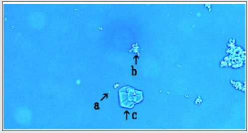
Microscopic appearance of DNA crown cell’s DNA crown cells using commercial human placenta DNA were prepared using adenosine-monolaurin with previously described methods [9]. A typical single DNA crown cell is shown in (Figure 1 arrow a). The cells were round and possessed cloud-like protrusions. They were approximately 3-4 μm in size, and many clusters in which DNA crown cells were gathered were also observed (Figure 1 arrow b). Crystal-like objects were also observed (Figure 1 arrow c), and DNA crown cells were observed within these objects. The crystallike objects may have been formed by Sph and DNA that were not used in the synthesis of DNA crown cells. Color was selected under microscopic observation to make it easier to observe the objects, but the color does not have a specific meaning, as described previously [8].
Formation of assemblies in mixtures of DNA crown cells with B. subtilis before incubation
B. subtilis was mixed with DNA crown cells, and immediately (before incubation) they were observed under a microscope. Many assemblies with different shapes and characteristics were observed (Figure 2, 3, and 4).
One assembly that is shown in (Figure 2) consists of three parts (arrows a, b, a nd c). Part a may consist of DNA crown cells and their clusters. Part b has a crystal-like appearance as shown in (Figure 1 arrow b). Part c appears to be a cluster of bacteria. DNA crown cells were also observed within the crystal. The sizes of the assemblies were not discussed, because the sizes of these assemblies may be not important, although they can be estimated from the size of a typical single cell (3-4μm).
The assembly shown in (Figure 3) may be constructed from parts a, b, and c, as shown in (Figure 2). It consists of a cluster like the DNA crown cells shown in part a, the crystal-like objects shown in part b, and the objects like bacteria in part c. The objects in part c resembled long bacteria. When bacteria are added to Sph-DNA, objects such as long bacteria are observed (unpublished data), so the objects in part c may be bacterial assemblies.
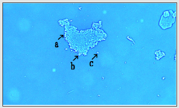
The assembly of clusters like DNA crown cells was observed, as shown in (Figure 4). Interestingly, a DNA crown cell was observed along the edge of the assembly (Figure 4 arrows a, c). However, it is unclear whether such phenomena are characteristic of DNA crown cells.
Figure 2: Assemblies in mixtures of DNA crown cells with Bacillus subtilis before incubation (1) One of these assemblies is shown in (Figure 2). The assembly consists of three parts (arrows a, b, and c). Part a may consist of DNA crown cells and their clusters. Part b shows a crystal-like appearance, as shown in (Figure 1) arrow b. A free DNA crown cell was also observed (arrow d). Part c has the appearance of many bacteria clustered together.
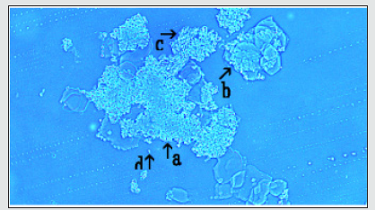
Figure 3: Assemblies in mixtures of DNA crown cells with Bacillus subtilis before incubation (2) The assembly shown in (Figure 3) may be constructed from parts a, b, and c, as shown in (Figure 2). The parts consist of a cluster of DNA crown cells in part a, crystal-like objects in part b, and bacteria-like objects in part c. A free bacterium was also observed (arrow d).
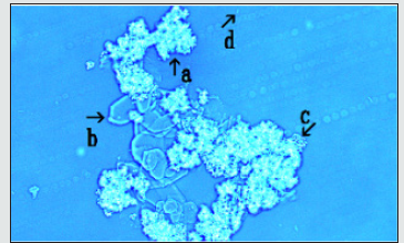
Figure 4: Assemblies in mixtures of DNA crown cells with Bacillus subtilis before incubation (3) The assembly of the cluster of DNA crown cells was observed, as shown in (Figure 4). Interestingly, each DNA crown cell was observed along the edge of the assembly (arrows a and b). A free DNA crown cell was also observed (arrow c).
Microscopic appearance after 3 h of incubation
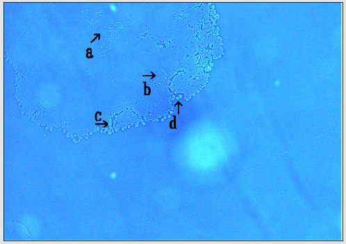
An assembly that was observed after 3 h of incubation is shown in (Figure 5). The assembly was flat, and many bacteria were observed within it (Figure 5 arrows, a, b). Objects of various shapes and sizes were also observed along the edge of the assembly. Objects (Figure 5 arrow c) appeared round before release from the assembly, and other objects (Figure 5 arrow d) appeared in a similar condition after release from the assembly. This suggests that round-like objects were generated from the assembly.
Figure 5: Microscopic appearance after 3 h of incubation the assembly was flat, and many bacteria were observed within it (arrows a and b). Objects of various shapes and sizes were also observed along the edge of the assembly. The objects (arrow c) that were round appeared before release from the assembly, whereas other objects (arrow d) appeared after release from the assembly.
Microscopic appearance after incubation for 24 h
The assemblies formed after incubation for 24 h are shown in (Figure 6, 7, 8, 9). The assembly shown in (Figure 6) was flat, and objects like bacteria were observed along its edge (Figure 6 arrows a, b, c, d). These objects along the edge were not observed when using E. coli DNA crown cells. These may be objects on which bacteria enclosed within the assembly grow. Based on careful microscopic observation, I found that DNA crown cell-like objects were bound to the tips of the bacteria-like objects (Figure 6, arrow d). Further observation will be needed to determine if the DNA crown cells influenced the bacteria within the assembly.
Figure 6: Microscopic appearance after incubation for 24 h (1) The assembly shown in Fig. 6 appears flat. Objects like bacteria were observed along the edge of the assembly (arrows a, b, c, and d), and a free bacterium was observed (arrow e).
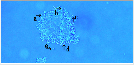
Figure 7: Microscopic appearance after incubation for 24 h (2) The assembly shown in Fig. 7 was flat, similar to that in Fig. 6, and objects like long bacteria were observed (arrows a and b). Afree DNA crown cell was also observed (arrow c).
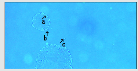
Figure 8: Microscopic appearance after incubation for 24 h (3) Objects like long bacteria separated from the assembly were also observed, as shown in Fig. 8 (arrow a). A free DNA crown cell was observed (arrow b).
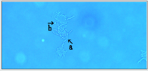
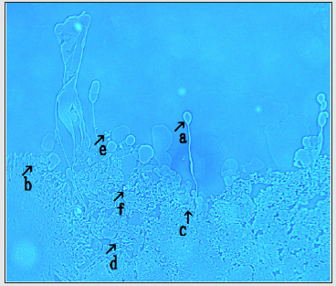
Figure 9: Microscopic appearance after incubation for 24 h (4) Objects of various sizes and shapes like balloon were observed (Fig. 9, arrow a), and small particles were observed within the assembly (arrows b and c). Objects like bacteria were also observed (arrow d), and free objects like DNA crown cells were observed inside (arrow f) and outside (arrow e) in the assembly.
The assembly shown in (Figure 7) was flat, like that shown in (Figure 6). However, the assembly possessed objects that resembled long bacteria (Figure 7 arrows a, b), and these may have been released from the assembly, as shown in (Figure 8 arrow a). As described above, it may be that DNA crown cells genetically influenced the bacteria. Moreover, the bacteria may have been physiologically influenced by the irregular growth environment, which increased their length.
A curious looking assembly is shown in (Figure 9), in which objects of various sizes and shapes like balloons were observed (Figure 9 arrow a). Small particles were also observed within the assembly (Figure 9 arrows b, c), which may serve as seeds on which objects grow like balloons. Bacteria-like objects were also observed (Figure 9 arrow d), and free-floating objects like DNA crown cells were observed inside (Figure 9 arrow f) and outside (Figure 9 arrow e) the assembly. As described previously [8], the observed assemblies consisted of Sph, DNA, Sph-DNA, DNA crown cells, and so on. Therefore, it was considered that the objects like balloon were also constructed from these components. In contrast, objects like balloon were also observed when using E. coli DNA crown cells (unpublished data), hence the objects like balloon may be not a characteristic of human placenta DNA crown cells. Presently, it is unclear whether objects like balloon are related to the generation of the bacteria-like objects or DNA crown cells.
Here, I demonstrated that the assemblies that were observed in the combination of E. coli DNA crown cells and B. subtilis were also observed in the combination of human placenta DNA crown cells with B. subtilis. In general, the assemblies that were formed appeared very similar. However, there were several differences based on microscopic observation. One difference is that crystal-like objects were observed in crown cells using human placenta DNA, as shown in (Figure 2,3) whereas the case which such objects were observed was rear in crown cells using E. coli DNA. This difference may depend on the components of the DNA. Sph-DNA, especially the Sph, binds various compounds as described previously [8], and compounds that form the crystal-like objects may be contained in human placenta DNA and not in E. coli DNA. However, it is unclear whether such objects have any functions within the assembly.
In addition, many bacteria-like objects were observed in assemblies formed with the use of human placenta DNA, as shown in (Figure 4), whereas a small number of bacteria-like objects were observed in assemblies formed with the use of E. coli DNA. The growth of bacteria enclosed within assemblies may not be suppressed with the use of placenta DNA, whereas it could be suppressed with the use of E. coli DNA. This suggests that there may be some differences in the quality of the assemblies depending on the kinds of DNA used for the preparation of DNA crown cells. Nevertheless, DNA crown cells were observed both with the use of E. coli DNA and human placenta DNA.
As described previously, DNA crown cells that were prepared in the present experiments multiplied within egg whites. Therefore, bacteria that were used to form the assemblies may not be necessary to form the DNA crown-like cells. It also remains unclear whether these objects were indeed DNA crown cells and were generated from the assembly.
In addition to whether DNA crown cells were generated within the assemblies, the fate of the bacteria enclosed within the assemblies also varied. In some cases, most of the bacteria were digested within the assemblies, but a small number of bacteria survived. In this case, it was considered that the compositions of the assemblies changed, and they contained new compounds. Such compounds may have been used for the generation of the DNA crown cell-like objects. In other cases, most of the bacteria remained alive within the assemblies. In this case, it was considered that the bacteria enclosed within each assembly were physiologically or genetically influenced within an environment unsuitable for bacterial growth, and as a result, bacterial transformation may have occurred.
Only B. subtilis were used in the present study. Therefore, in future studies, we should investigate assemblies formed by a combination of DNA crown cells with different types of bacteria. This will allow us to evaluate the scientific meaning of the assemblies formed between DNA crown cells and bacteria. However, even with the bacteria used in the present experiments, it was notable that it was carried out using commercially available reagents through a series of experiments including the preparation of DNA crown cells, and the methods by which the DNA crown cells were prepared have already been established [1,2,9].
Therefore, the present system may serve as an experimental platform to resolve difficult subjects that have been discussed for a long time in the life sciences, such as the origin of life, cells, bacteria, plasmids, and the living status of viruses, and may have industrial applications.
References
- Inooka S (2012) Preparation and cultivation of artificial cells. App. Cell Biol 25: 13-18.
- Inooka S (2016) Preparation of Artificial Cells Using Eggs with Sphingosine-DNA. J Chem Eng Process Technol 7(1): 277.
- Inooka S (2016) Aggregation of sphingosine-DNA and cell construction using components from egg white. Integrative Molecular Medicine 3(6): 1-5.
- Inooka S (2017) Systematic Preparation of Bovine Meat DNA Crown Cells App. Cell Biol 30: 13-16.
- Inooka S (2018) Systematic Preparation of DNA (Akoya pearl oyster) Crown Cells App. Cell Biol 21-34.
- Inooka S (2013) Preparation of Artificial Cells for Yogurt Production App. Cell Biol. 26: 13-17.
- Inooka S (2014) Preparation of Artificial Human Placental Cells App. Cell Biol 27: 43-49.
- Inooka S The assembly formation by co-cultures of DNA (Escherichia coli) crown cells with Bacillus subtillus cells. Journal of Biotechnology and Bioresearch In press.
- Inooka S (2017) Biotechnical and Systematic Preparation of Artificial Cells (DNA Crown Cells). The Global Journal of Research in Engineering 17(1-c).

Top Editors
-

Mark E Smith
Bio chemistry
University of Texas Medical Branch, USA -

Lawrence A Presley
Department of Criminal Justice
Liberty University, USA -

Thomas W Miller
Department of Psychiatry
University of Kentucky, USA -

Gjumrakch Aliev
Department of Medicine
Gally International Biomedical Research & Consulting LLC, USA -

Christopher Bryant
Department of Urbanisation and Agricultural
Montreal university, USA -

Robert William Frare
Oral & Maxillofacial Pathology
New York University, USA -

Rudolph Modesto Navari
Gastroenterology and Hepatology
University of Alabama, UK -

Andrew Hague
Department of Medicine
Universities of Bradford, UK -

George Gregory Buttigieg
Maltese College of Obstetrics and Gynaecology, Europe -

Chen-Hsiung Yeh
Oncology
Circulogene Theranostics, England -
.png)
Emilio Bucio-Carrillo
Radiation Chemistry
National University of Mexico, USA -
.jpg)
Casey J Grenier
Analytical Chemistry
Wentworth Institute of Technology, USA -
Hany Atalah
Minimally Invasive Surgery
Mercer University school of Medicine, USA -

Abu-Hussein Muhamad
Pediatric Dentistry
University of Athens , Greece

The annual scholar awards from Lupine Publishers honor a selected number Read More...


