Lupine Publishers Group
Lupine Publishers
Menu
ISSN: 2690-5752
Short Communication(ISSN: 2690-5752) 
Unraveling the Canvas Employed by Italian Painter Carlo Ferrario in Large-Format Artworks at the National Theater of Costa Rica Volume 4 - Issue 3
MD Barrantes Madrigal1,2, Paula Calderón Mesén3, Mariela Agüero Barrantes4 and OA Herrera Sancho2,3,5,6*
- 1Escuela de Química, Universidad de Costa Rica, 2060 San Pedro, San José, Costa Rica
- 2Instituto de Investigaciones en Arte, Universidad de Costa Rica, 2060 San Pedro, San José, Costa Rica
- 3Centro de Investigación en Estructuras Microscópicas, Universidad de Costa Rica, 2060 San Pedro, San José, Costa Rica
- 4Museos del Banco Central de Costa Rica, 10104 San José, Costa Rica
- 5Escuela de Física, Universidad de Costa Rica, 2060 San Pedro, San José, Costa Rica
- 6Centro de Investigación en Ciencias Atómicas Nucleares y Moleculares, Universidad de Costa Rica, 2060 San Pedro, San José, Costa Rica
Received:July 02, 2021 Published: July 12, 2021
Corresponding author:OA Herrera Sancho, Instituto de Investigaciones en Arte, Universidad de Costa Rica, 2060 San Pedro, San José, Costa Rica
DOI: 10.32474/JAAS.2021.04.000190
Abstract
The comprehensive study of artworks is key to understanding the temporal evolution of all the factors that surround it. A painting is generally composed of multiple layers and materials which are hidden from the naked eye because their surface colors are highly captivating. However, a fundamental pillar of an artwork is the canvas which usually has a special preparation to obtain the final finish desired by the artist. Here, we study the canvas employed by Italian painter Carlo Ferrario in two large-format paintings at the National Theater of Costa Rica. The main goal of the current research was to determine the state of conservation and major materials of the canvases used by the artist. We systematically explored samples of these canvases by means of Optical Microscopy, Scanning Electron Microscopy with Energy Dispersive X-ray Spectroscopy and Fourier Transform Infrared spectroscopy. We were able to identify the canvas as, potentially, hemp based on morphological characteristics of the fiber.
We estimated the amount of fatty acids by means of Gas Chromatography Mass Spectroscopy resulting primarily in suberic, azelaic, sebasic, palmitic (C16), and stearic (C18). Hence, it could conceivably be hypothesised that the pictorial method in the studied paintings corresponds to the oil technique. This combination of findings provides support to establish efficient methodologies to the diagnosis of the conservation state of canvases. Furthermore, to comprehend that preserving these works of art for future generations as part of our Costa Rican cultural heritage demands a deeper understanding of the paintings, especially given that art conservation in the tropics is a topic still largely unexplored.
Keywords: Canvas; Hemp; Analytical Chemistry; Characterization and Analytical Techniques; Cultural Heritage
Introduction
Art canvases are the backbone of every artwork but are often set aside when analyzing all the different layers and elements of a painting. The fibers that integrate the canvas have their own physical and chemical properties, which can affect the overall condition of the object [1]. Understanding the fibers can give valuable information about the historical background, such as the technology used, geographical location, and possible elements ingrained in the weave. Furthermore, by identifying the fiber within the canvas, feasible projections about its behavior can be created, and therefore, conservation treatments can be proposed [2]. Located at the National Theater of Costa Rica (NTCR), specifically in the former men’s canteen, Musas I and Musas II are two largeformat artworks situated at the ceiling. Both paintings are located side by side, measuring 2.96m x 6.17m (WxH) each. They both have been previously studied, revealing first-hand results from an interdisciplinary scientific approach [3]. With these new results, the objective is to emphasize the importance of the fibers and how these elements of the painting can tell a parallel story. The samples were extracted from damaged areas of the painting; therefore, they have particular shapes and small sizes. The specimens encompass fibers, the first ground layer, pigments used, and the binder. Fiber samplings can be described as individual threads splintering from the core weave structure, possibly due to its condition. The remaining part of the paper proceeds as follows: Section II describes the methodology used for the morphological and chemical composition of the canvas and binder. Following this, in the Results section, we carry out a discussion of the most important findings of the studied canvas of approximately 125 years. Finally, we finish with concluding remarks and next experimental steps to provide a comprehensive diagnostic of the state of conservation of the canvas.
Methodology
Morphological characterization of canvas fibers
Canvas’ fibers samples were characterized by optical microscopy Olympus IX51 and scanning electron microscopy Hitachi S-3700N with energy dispersive x-ray spectroscopy detector IXRF Systems (SEM-EDX) (primary voltage, 20 keV; working distance, 10-12 mm). Elemental composition was obtained under variable pressure conditions. Samples were covered with gold for image acquisition.
Chemical composition of the canvas and binder
Fourier Transform Infrared spectroscopy with Attenuated Total Reflectance (FTIR-ATR)
In order to identify the composition of the canvas FTIR-ATR analysis was carried out. A Scientific Evolution Nicolet 6700® spectrometer was used for performing ATR measurements at one thread extracted from the canvas. Spectra were obtained using transmittance mode. Data was acquired in the range of 4000 to 650 cm−1, with 1 cm−1 spectral resolution.
Gas Chromatography Mass Spectroscopy (GC-MS)
GC-MS was performed for identifying the binder of these artworks, according to the procedure used by Alcántara-García, J. and Nix, M. (2018) [4]. The sample analyzed included a fragment of the painting layers (about 1 mm2) with few strands of the canvas (about 1 cm). Samples were treated with Grace Alltech MethPrep II reagent in benzene (≤100 μL of 1:2), using Thermo Fisher Scientific autosampler vials, to transform any carboxylic acids or esters into their methyl ester derivatives. The vials were heated at 60°C in a Lab-Line Multi-Blok heater; after one hour, the vials were removed and cooled. A sample volume of 1μL was injected onto an Agilent Technologies 7820 gas chromatograph equipped with a HP-5MS column (30m × 250μm × 0.25μm of film thickness, 5% phenyl methyl siloxane, flow rate of 1.5 mL/minute), an Agilent 5975 mass selective detector (MSD) and an automatic liquid injector. The inlet temperature was 320°C (splitless mode) with a nine-minute solvent delay. The GC oven temperature program was 55°C for 2 minutes, then ramped at 10°C/minute to 325°C, and 10 minutes of isothermal period. The transfer line temperature to the MSD (SCAN mode) was 280°C, the source at 230°C and the MS quad at 150°C. These experimental conditions were programmed and controlled by the Agilent Technologies G1701EA GC/MSD Chem Station Control software. The data was recorded on Agilent MSD Enhanced Chemstation Data Analysis software with NIST MS Search 2.0 database, they also facilitated the chromatograms and mass spectra interpretation.
Results and Discussion
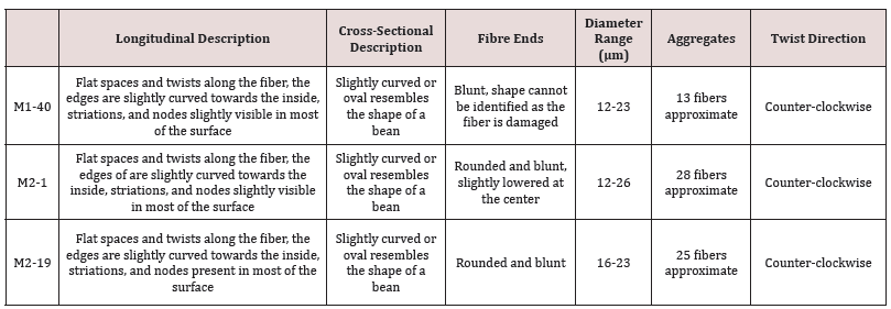
During the 19th century, the most common canvas fibers in Europe were hemp, linen, a combination of both or linen and cotton; therefore, a thorough fiber identification was necessary to confirm the fiber supporting the painting [5]. Based on SEM fiber analysis images and optical microscopy the fiber in the canvas most likely corresponds to hemp. Table 1 describes the visible/physical features of the analyzed samples. The features underlined are suggested by Carr et al. (2008) [1] and Nayak et al. (2020) [6] as inherent pointers of a specific fiber. The main difference between hemp and linen, the other possible option based on historical accuracy, is the prominent nodes present in linen but less prominent in hemp combined with striations [7]. Hemp fibers have slight twists similar to cotton and an overall irregular surface [8]. The twist on the hemp fibers showed to be counterclockwise according to SEM microscopy images [1]. Figure 1 exemplifies the described morphological features visible in the hemp fibers. All SEM and optical microscope images belong to sample M1-40 see Figure 2 in Barrantes, et al. in order to find the grid location of this sample [3]. Optical microscope images evidence the tiny nodes and striations that compose the fiber as well as its irregular surface. CAMEO materials database [9] images emphasize these features as hemp exclusive. Aside from all the mentioned characteristics, SEM image 1.c shows a slightly curved edge towards the inside, forming a centerline in the fiber [1,10]. The morphological aspects of the fiber are accentuated with SEM-EDX and FTIR analyses. SEM-EDX analysis of fiber samples of Musas I and Musas II paintings are presented in Figure 2. The EDX analysis showed the most representative elements presented on the sample, with carbon and oxygen with the highest concentration. These elements are constituents of cellulose and hemicellulose, main components of the hemp [6]. The fibers also have crystals particles (Figure 2c) corresponding to pigments and material that remains which was probably used by the artist to prepare the initial stages of the canvas. The largest and brightest crystals correspond to lead, as shown in the EDX mapping. The main layers of Musas I and Musas II have been defined by Barrantes et al. (2021) [3], showing pictorial layers with a color palette of lead red, ultramarine blue, vermilion, viridian and chrome yellow and lead white and zinc white. Lead red and lead white, have been recommended for protecting the back of a canvas [11,12]. Calcium is present on the first ground layer in the form of calcium carbonate known as chalk, which was commonly used as white pigments in antiquity [13] and ground material [12,14]. According to previous studies of this artwork [3], lead is present in the second ground layer and calcium in the first ground layer. Hemp is a plant fiber obtained from Cannabis sativa L. and is constituted by 67-80 % cellulose [1].
The presence of cellulose is verified by a preliminary analysis of a canvas sample by Fourier Transform Infrared Spectroscopy with Attenuated Total Reflectance (FTIR-ATR). Cellulose is a natural polymer whose structural monomer is a 1-4 linked β-D-glucose [15]. It means a six atoms heterocycle of carbons and oxygen, with hydroxyl groups bonded to the cycle carbon [15]. Those groups contribute to the formation of intermolecular hydrogen bonds and provide the mechanical properties of this macromolecule [15]. Figure 3a indicates the characteristic signals of this compound. First, O-H stretching (near to 3300 cm-1) evidence the hydroxyl groups. Later, hydrocarbon asymmetric (2917 cm-1) and symmetric and C-H stretching vibrations (2850 cm-1).Additionally, signals indicated as 1 correspond to O-H and OCH bending vibrations (1440 cm-1) and signals 2 show C-O-C asymmetric stretching (at 1160 cm-1). Those peaks are observed because of the hydroxyl and ether oxygens in the compound (Fan et al., 2012). Other important bands observed, pointed out as 3, are the C-C, C-OH, C-H ring and side group vibrations (at 1050 – 990 cm-1) because of the cyclic structure of cellulose [10].
Figure 1: Morphological characteristics of canvas’s fibres. a) & b) optical microscope view, SEM details of c) longitudinal description, d) cross-sectional description, e) fibre ends, f) diameters, g) aggregates and h) twist direction.
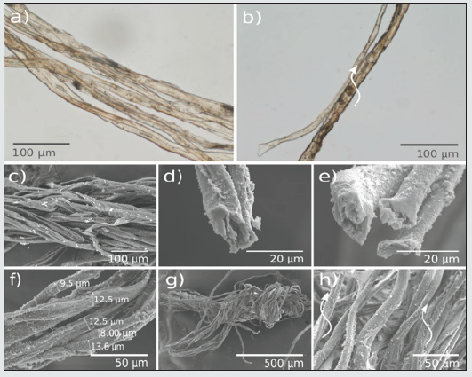
Figure 2: SEM-EDX (scanning electron microscopy energy-dispersive X-ray spectroscopy) of hemp’ fibers: (a) Musas I painting and (b) Musas II painting. (c) Fibers and crystal’s micrographs details, showing as brighting particles over the fibers and EDX map on Musas II painting.
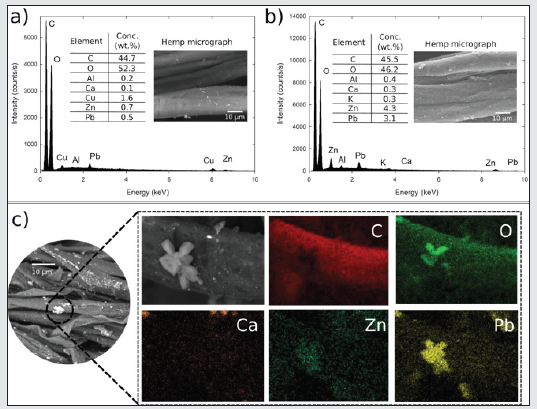
Figure 3: Spectroscopic characterization of materials. a) FTIR-ATR spectrum of canvas’ fibre showing O-H stretching, C-H asymmetric (2917 cm-1) and symmetric stretching, C=O stretching vibration, O-H, HCH and OCH bending vibration (indicated as 1), C-O-C asymmetrical stretching (2), C-C, C-OH, C-H ring and side group vibrations (3). b) Chromatogram of painting sample showing signals of dicarboxylic acids: suberic, azelaic, sebacic, palmitic and stearic acids.
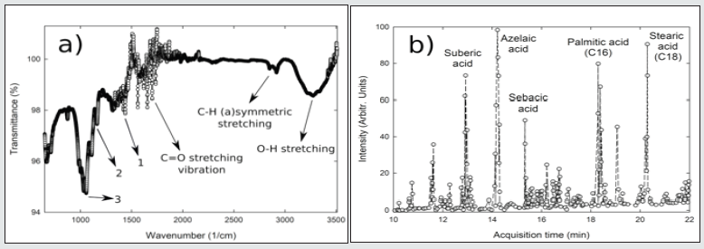
A sample of a thread of canvas impregnated with pictorial layers of Musas I was analyzed by GC-MS. Significant signals of characteristic fatty acids in drying oil appear in the chromatogram of Figure 3.b. The most important acids found were suberic, azelaic, sebasic, palmitic (C16) and stearic (C18) acids. These results reveal information about the binder of the painting. The most common drying oils are linseed poppy seed, and walnut oils [12]. The quantities of those acids found in paintings is quite diverse because it depends on the type of drying oil the artists used and the aging processes in the painting [16]. The ratio of palmitic to stearic acid (C16/C18) is commonly used for identification of drying oils. In the present study it corresponds to 1.09. According to the classification established by Manzano (2011) [16], this is a characteristic value for linseed oil binder. On the other hand, due to the presence of drying oil, we suggest that the pictorial technique in the studied paintings corresponds to the oil technique. These findings focused on Musas I and Musas II contrast with the pictorial technique of other artists at the NTCR. For instance the egg yolk in the Main Curtain, studied by Morice, et.al (2019) [17], indicates tempera as its pictorial technique. In general, it is key to separate all the components of an artwork, such as canva’s fibers, binder, and pigments for research purposes. Fibers contribute to both support and the preservation of the painting, therefore a continuous study is necessary. Since fiber samples were splintering from the core weave, we can highlight that the degradation of cellulose by chemical [18,19] and biological [3] sources is a process that contributes to the changes of the condition of artworks over time. Henceforth, a thoughtful preservation strategy is needed for future instances.
Conclusion
This study contributes to the knowledge about pigments, materials, and canvas’ fibers used by artists and the degradation process that occurs in neotropical regions. This first approach led us to study the canvas’ fibers used in the National Theater Musas I and Musas II paintings and classified the textile as hemp through morphological and chemical characterization. The next step is to understand the degradation process of hemp. Therefore, pigments and ground materials need to be tested and analyzed. Multidisciplinary studies about Costa Rican paintings can shed light on discoveries about historical contexts and cultural heritage and contribute to the field of conservation. In essence preserving artworks, especially in tropical countries, has a different set of challenges which require a deeper analysis and understanding.
Acknowledgements
We would like to thank Jocelyn Alcántara-García from University of Delaware for all the valuable assistance in GC-MS measurements. We give special thanks to Centro de Electroquímica y Energía Química and Centro de Investigación en Estructuras Microscópicas for allowing us to perform various measurements, specifically FTIR-ATR and SEM-EDX, respectively. We want to acknowledge the support of all the staff of the National Theater of Costa Rica for all the logistics and making it possible to carry out this study and especially to our friend Carmen Marín-Cruz who is no longer with us. We are grateful for the support given by the Vicerrectoría de Investigación at the Universidad de Costa Rica to carry out this research work.
Discussion and Conclusion
The highest subjective test results are Joy and Sadness. They represent the greatest variation between human perceptions and ML. These could be further used in sentiment polarity, which could support mass sensing in any market sensing product. The scores in Human Neutral column spread over ML emotion rows. This could mean that some levels of emotion could be identified as neutral. In this preliminary study, a Thai short expressive message corpus was created from Twitter. The emoji usage in them indicates emotions. A set of 22 emotional emoji groups used in a Thai context were formed by using K-nearest [16,17]. An analysis of these groups suggests that the emotions portrayed could be related to Deep Moji’s architecture which could provide a possible list of emojis relating to a short text message input. Clustering the multi-label emoji groups according to Ekman’s 6 basic emotions can be used to interpret the social emotional meaning of the message. The emojis in Table 2 are derived from the groups in Table 1. They are used in a final automatic social emotion detection system. It is a scheduled crawler for up-to-date Thai tweets and passes them to an emoji classification which leads to a group of emotions. Thus, a demo prototype called Emo Sense can be established. http://pop.ssense.in.th/EmoSense/.
References
- Carr D, Cruthers N, Smith C, Myers T (2008) Identification of selected vegetable textile fibres. Studies in Conservation 53(sup 2): 75-87.
- Conejo Barboza G, Libby E, Marín C, Herrera Sancho OA (2020) Discovery of Vespasiano Bignami paintings at the National Theater of Costa Rica through technical photography and UV-Vis spectroscopy. Heritage Science 8(125): 1-10.
- Barrantes Madrigal MD, Zúñiga Salas T, Arce Tucker RE, Chavarría Sibaja A, Sánchez Solís J, et al (2021) Revealing time’s secrets at the National Theatre of Costa Rica: Innovative software for cultural heritage research. Scientific Reports 11(8560): 1-16.
- Alcántara García J, Nix M (2018) Multi-instrumental approach with archival research to study the Norwich textile industry in the late eighteenth and early nineteenth centuries: the example of a Norwich pattern book dated c. 1790–1793. Heritage Science 6(1): 1-15.
- Vanderlip de Carbonnel K (1980) A Study of French Painting Canvases. Journal of the American Institute for Conservation 20(1): 3-20.
- Nayak R, Houshyar S, Khandual A, Padhye R, Fergusson S (2020) Identification of natural textile fibres. En Handbook of Natural Fibres: Second Edition (Second Vol. 1) pp. 503-534.
- Boerma F, Brokerhof A, Van Der Berg S, Tegelaers J (2013) Unraveling Textiles. A handbook for the preservation of textile collections. Archetype Publications Ltd, London, UK.
- Rahman M, Sayed Esfahani M (1979) Study of Surface Characteristics of Hemp Fibres Using Scanning Electron Microscopy. Indian Journal of Fibre & Textile Research 4(3): 115-120.
- CAMEO (2017) CAMEO Materials Database.
- Lu N, Swan RH, Ferguson I (2012) Composition, structure, and mechanical properties of hemp fiber reinforced composite with recycled high-density polyethylene matrix. Journal of Composite Materials 46(16): 1915–1924.
- Feller RL (Ed.) (1986) Artist´s Pigments: A Handbook of their History and Characteristics Volume 1. Washington, USA.
- Mayer R (1970) The Artist’s Handbook of Materials and Techniques (Third Edit) New York: The Viking Press, Inc.
- Goffer Z (2007) Archaeological Chemistry (2nd ed.) John Wiley & Sons, Inc, USA.
- Stols Witlox M, Ormsby B, Gottsegen M (2018) Grounds 1400-1900. En J H Stoner & R. Rushfield (Eds.) Conservation of easel paintings (First Edit.) pp. 161-188.
- Fan M, Dai D, Huang B (2012) Fourier Transform Infrared Spectroscopy for Natural Fibres. En S. Salih (Ed.) Fourier Transform - Materials Analysis (1st Ed.) pp. 45-68.
- Manzano E, Rodriguez Simón LR, Navas N, Checa Moreno R, Romero Gámez M, et al (2011) Study of the GC-MS determination of the palmitic-stearic acid ratio for the characterisation of drying oil in painting: La Encarnación by Alonso Cano as a case study. Talanta 84(4): 1148-1154.
- Morice JG, Benavides Rodríguez J, Conejo Barboza G, Marín C, Montero M L, et al (2019) A Brief Insight Into the Secrets of the 120-Year-Old Main Curtain of the National Theatre of Costa Rica Through Non-Destructive Characterization Techniques. Journal of Conservation and Museum Studies 17(1).
- Bitossi G, Giorgi R, Mauro M, Salvadori B, Dei L (2005) Spectroscopic techniques in cultural heritage conservation: A survey. Applied Spectroscopy Reviews 40(3): 187-228.
- Oriola M, Možir A, Garside P, Campo G, Nualart Torroja A, et al. (2014) Looking beneath Dalí’s paint: Non-destructive canvas analysis. Analytical Methods 6(1): 86-96.

Top Editors
-

Mark E Smith
Bio chemistry
University of Texas Medical Branch, USA -

Lawrence A Presley
Department of Criminal Justice
Liberty University, USA -

Thomas W Miller
Department of Psychiatry
University of Kentucky, USA -

Gjumrakch Aliev
Department of Medicine
Gally International Biomedical Research & Consulting LLC, USA -

Christopher Bryant
Department of Urbanisation and Agricultural
Montreal university, USA -

Robert William Frare
Oral & Maxillofacial Pathology
New York University, USA -

Rudolph Modesto Navari
Gastroenterology and Hepatology
University of Alabama, UK -

Andrew Hague
Department of Medicine
Universities of Bradford, UK -

George Gregory Buttigieg
Maltese College of Obstetrics and Gynaecology, Europe -

Chen-Hsiung Yeh
Oncology
Circulogene Theranostics, England -
.png)
Emilio Bucio-Carrillo
Radiation Chemistry
National University of Mexico, USA -
.jpg)
Casey J Grenier
Analytical Chemistry
Wentworth Institute of Technology, USA -
Hany Atalah
Minimally Invasive Surgery
Mercer University school of Medicine, USA -

Abu-Hussein Muhamad
Pediatric Dentistry
University of Athens , Greece

The annual scholar awards from Lupine Publishers honor a selected number Read More...


