Lupine Publishers Group
Lupine Publishers
Menu
ISSN: 2690-5752
Short Communication(ISSN: 2690-5752) 
Herculaneum 79 AD: Neuronal Tissue Preservation from a Vitrified Human Brain Volume 4 - Issue 2
Pierpaolo Petrone1*, Vincenzo Graziano1, Giuseppe Castaldo2, Mariano Paternoster1, Francesco Sirano3, Guido Giordano4, Alessandra Pensa4, Alessandro Vona4, Emanuele Capasso1 and Massimo Niola1
- 1Università degli Studi di Napoli Federico II, via Pansini 5, Naples, Italy
- 2CEINGE - Biotecnologie Avanzate. Via Gaetano Salvatore 486, Naples, Italy
- 3Parco Archeologico di Ercolano, Corso Resina 187, Naples, Italy
- 4Università degli Studi Roma Tre, Largo San Leonardo Murialdo 1, Rome, Italy
Received:May 25, 2021; Published: June 02, 2021
Corresponding author:Pierpaolo Petrone, Università degli Studi di Napoli Federico II, Naples, Italy
DOI: 10.32474/JAAS.2021.04.000184
Abstract
In AD 79 the town of Herculaneum was suddenly hit and overwhelmed by successive volcanic ash-avalanches, fast moving clouds of hot volcanic ash and gases, capable of killing all residents who were not yet evacuated. The scientific studies on the Herculaneum victims are now standing from the first discovery in the early 1980s of hundreds of skeletons of people crowding the beach and a series of waterfront chambers, fixated into a final vital stance by the first of the deadly incoming pyroclastic currents. Multidisciplinary studies on the victims’ skeletons and their biogeoarchaeological context shed light on the dynamic impacts of the 79 AD Plinian eruption on the area around the volcano and on the causes of death of its inhabitants. A recent unprecedented archaeological discovery revealed unique evidence of preservation of a vitrified brain from a human victim found in the town. SEM analysis of brain and spinal cord vitrified remains showed an integrally preserved central nervous system. Results from site research combined with lab analysis offer new insights concerning the unique conditions occurred during the 79 AD Vesuvius eruption, with crucial implications for the present-day risk of a similar outcome to around three million people living close to the volcano, including metropolitan Naples.
Keywords:Forensic Anthropology; Bioarchaeology; 79 AD Eruption Victims; Vesuvius
Introduction
Up to now, there have been sporadic discoveries of wellpreserved cerebral tissue from ancient human remains [1]. Under certain taphonomic conditions that prevent soft tissue decomposition, these remains are typically saponified [2]. However, ancient brains reported in literature show only partial preservation of neuronal structures [3]. During our recent paleoforensic survey at the archaeological site of Herculaneum, we discovered glassy material within the cranial cavity from a victim of the 79 AD Vesuvius eruption, apparently derived from his brain. Recent examination by proteomic analysis and gas chromatography-mass spectrometry of this material allowed us to identify fatty acids of human hair fat and several proteins of human brain origin, thus indicating preservation of vitrified human brain tissue [4]. The conversion of human tissue to glass (vitrification) occurred due to the rapid cooling of the volcanic ash deposit after exposure to the burning ash cloud [5]. Previous heating bone experiments showed temperatures of about 500 °C [6], as also confirmed by our recent reflectance analysis on carbonized wood from Herculaneum [4]. Recently, examination of the ultrastructure of vitrified human remains showed several typical features of the central nervous system (CNS) from brain and spinal cord tissue [7].
Historical Background
In AD 79, a major explosive eruption hit by hot ash-avalanches the Vesuvius towns up to 20 kilometres away, causing thousands of fatalities [8]. Herculaneum, located 6 kilometres from the volcano, was rapidly buried beneath 20 metres thick volcanic deposits. The environmental burial conditions ensured the town to be preserved intact until the first discovery of its theatre, in 1710. Some decades later, during the early Bourbon exploration, the first human victims were discovered on November 18, 1739. A whole urban settlement gradually revealed itself as it had been buried by volcanic ashes and returned in its integrity. However, most exceptional has been the discovery of some hundreds of human victims, unearthed in several seafront chambers and on the beach during archaeological investigations in the second half of the 1900s [9]. The volcanic deposits have preserved intact for centuries the corpses of these victims as time capsules, a source of unique bio-anthropological information [10].
The guardian of the collegium augustalium
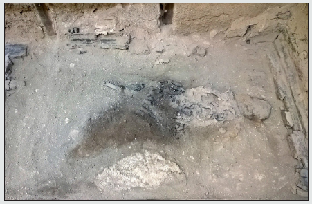
Figure 1: A complete overview of the 79 AD eruption’s victim. The original context of discovery (Collegium Augustalium, Herculaneum).

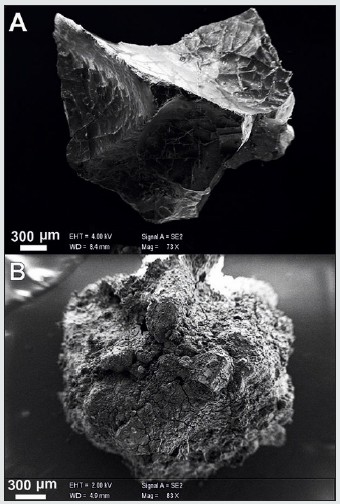
Figure 2: Vitrified human tissues from the central nervous system. A. brain fragment; B. spinal cord fragment (SEM, scale bars in micron).

In the mid-1960s, during the archaeological excavations of the town, in the Collegium Augustalium a small room was discovered containing a human victim lying on a wooden bed. This young adult male is believed to have been the guardian of the College, a centre of the cult of the Emperor Augustus. The victim is lying ventrally, face down in the volcanic ash (Figure 1). The skull and the postcranial bones show complete charring and cracking induced by the hot pyroclastic ash surge [11], a high-speed turbulent cloud rich in hot gases, ash, and steam [12].
In a recent survey at the archaeological site we identified vitrified cerebral tissue within the skull of this 79 AD eruption victim [4]. Microspectroscopic analysis by Raman technique has shown this tissue to consist exclusively of organic matter. We then carried out a scanning electron microscope (SEM) investigation on the vitrified tissue collected from the skull and the spinal cord (Figure 2A). Examination of the vitrified ultrastructure revealed several features, typical of the central nervous system (CNS) (Figure 2B). This type of remains is unique for the excellent quality of the tissue preservation. In literature several cases of ancient brains are reported which are preserved due to natural mummification or saponification, which allowed only partial preservation of neuronal brain tissue [1-3]. At Herculaneum, instead, the structures have been immobilized in their native condition, as a result of a natural process of vitrification induced by the local environmental conditions during the eruption [4,5].
Neuronal ultrastructures
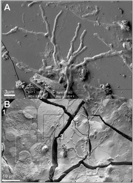
Figure 3: Structures of the central nervous system. A. SEM image of brain axons; B. SEM image of spinal cord axons intercepting cell bodies and sheath-shaped structures (scale bars in micron).

According to our SEM investigation, distinctive result of the CNS tissue vitrification has been the preservation of a system of axonlike tubular structures running across the cerebral matrix (Figure 3A) [7]. The observed neuronal architecture is analogous to that one detectable from biological samples after manual drop plunging protocol, or high-pressure freezing protocol followed by freezefracturing and cryo-SEM imaging [13]. These structures are mostly elongated and round in shape, as described in literature [14]. The mean diameter ranges between 550 and 830 nm, as expected for white matter axons which are in the medium 0·5 - 0·8 μm size range [15]. Such tubular structures are smaller in diameter than blood vessels in the cerebrovascular system [16]. A complex neuronal system was also identified in a glassy residue of spinal cord from the back bone. A basic structure is recurrent within the vitrified matrix: the neuronal architecture shows the preservation of a number of cellular bodies interconnected by a reticulum of multiple tubular structures (Figure 3B), whose morphology and size are analogous to those of neurons and axons. An image analysis procedure was used to extract quantitative information from SEM images. The round-shaped cell bodies, mean sized from 2·7 to 14·2 μm, show a cell membrane and an intracellular lumen filled by filamentous structures and nanovesicles. The sections of the neuronal cell body were classified in three groups depending on diameter (μm, mean ± SEM) (14·17 ± 1·66; 8·06 ± 0·32; 2·72 ± 0·28) and mean area (μm2, mean ± SEM) (157·9 ± 36·2; 53·2 ± 6·8; 7v1 ± 1·1).
We have also calculated the mean diameter (nm, mean ± SEM) of 15 brain axons (717·7 ± 24·0) and 15 spinal cord axons (672·1 ± 20·2), values that are comparable to those of white matter axons.20 Furthermore, the free axons that we have identified in the vitrified brain tissue (Figure 3A) have proved to possess the typical myelin periodicity as resulting from neural network processing based on our specific mathematical method elaborated through MATLAB software [17]. Through this method, in two different vitrified brain axons we detected several different membrane layers that wrapped around individual axons and formed compact myelin: the pattern alternating darker lines and white spaces is the same as seen in vivo for mammalian CNS myelin [18]. In contrast with myelinated axons observed in the cerebral tissue, axons in continuity with cells detected from the spinal cord appear as non-myelinated, and characterized by a smaller mean diameter (452·5 ± 14·5). We have also identified the presence of regular nanotubes inside the preserved cytoplasmic matrix of a neuronal cell body, as evidenced by our algorithm image processing [6]. The mean diameter of such nanostructures is of about 23 nm, value which is analogous to microtubules size [19]. Through the same procedure we discovered that the CNS matrix of both the brain and spinal cord is made up of recurring, similar nanostructures. This evidence suggests that the preservation process of neuronal tissue induced by vitrification is the same for all the CNS structural components. On the base of the ultrastructural features that we observed on archaeological vitrified human tissue, we have classified the cell bodies as neurons and the axon-like processes as axons. We hypothesize that the unique natural process of vitrification occurred during the 79 AD eruption has locked the structure of the CNS, thus preserving intact its morphology. The detection of original neuronal structures of the central nervous system indicates the durable preservation of human brain and spinal cord ancient tissues.
Acknowledgment
This study was not supported by any funding source. The corresponding author (P.P.) confirm that he had full access to all data in the study and had final responsibility for the decision to submit for publication. We thank the archaeological staff of the Archaeological Park of Herculaneum for their help in the field. We would also thank Viola Desiato for her help in bibliographical research.
Author Contributions
P.P. designed and supervised the project; P.P. and F.S. designed and carried out the archaeological investigation; P.P., F.S., G.G., A.P. and A.V. performed data curation and formal analysis; P.P., V.G., G.C., M.P., F.S., G.G., A.P., A.V., E.C. and M.N. wrote the manuscript.
Competing Interests
The authors declare no competing interests.
References
- Papageorgopoulou C, Rentsch K, Raghavan M, Maria Ines Hofmann, Giovanni Colacicco, et al. (2010) Preservation of cell structures in a medieval infant brain: A paleohistological, paleogenetic, radiological and physico-chemical study. NeuroImage 50(3) : 893-901.
- Ubelaker DH and Zarenko KM (2011) Adipocere: What is known after over two centuries of research. Forensic Sci Int 208(1-3): 167-172.
- Doran GH, Dickel DN, Ballinger Jr WE, O F Agee, P J Laipis, et al. (1986) Anatomical, cellular and molecular analysis of 8,000-yr-old human brain tissue from the Windover archaeological site. Nature 323(6091) : 803-806.
- Petrone P, Pucci P, Niola M, Peter J Baxter, Carolina Fontanarosa, et al. (2020) Heat-induced brain vitrification from the 79 AD Vesuvius eruption. New Engl J Med 382(4): 383-384.
- Giordano G, Zanella E, Trolese M, Baffioni C, Vona A, et al. (2018) Thermal interactions of the AD79 Vesuvius pyroclastic density currents and their deposits at Villa dei Papiri (Herculaneum archaeological site, Italy). Earth Planetary Science Letters 490 : 180-192.
- Petrone P, Giordano G., Vezzoli E, Alessandra Pensa, Giuseppe Castaldo, et al. (2020) Preservation of neurons in an AD 79 vitrified human brain. PLoS One 15(10): e024001.
- Mastrolorenzo G, Petrone P, Pappalardo L, FM Guarino (2010) Lethal thermal impact at the periphery of pyroclastic surges: evidences at Pompeii. PloS One 5(6): e11127/1-12.
- Petrone P (2019) The Herculaneum victims of the 79 AD Vesuvius eruption: a review. J Anthropol Sci 96: 69-89.
- Mastrolorenzo G, Petrone P, Pagano M, Incoronato A, Baxter PJ,et al. (2001) Herculaneum victims of Vesuvius in AD 79. Nature 410(6830) : 769-770.
- Petrone P (2011) Human corpses as time capsules: new perspectives in the study of past mass disasters. J Anthropol Sci 89: 3-6.
- Petrone P, Pucci P, Vergara A, Amoresano A, Birolo L, et al. (2018) A hypothesis of sudden body fluid vaporization in the 79 AD victims of Vesuvius. PloS One 13(9): e0203210.
- Mastrolorenzo G, Petrone P, Pappalardo L and Sheridan MF (2006) The Avellino 3780-yr-B.P. catastrophe as a worst-case scenario for a future eruption at Vesuvius. Proc Nat Acad Sci USA 103(12): 4366-4370.
- Nakatomi R, Hayashida T, Fujimoto K (2005) Cryo-SEM and subsequent TEM examinations of identical neural tissue specimen. Brain Res Prot 14(2): 100-106.
- Abdollahzadeh A, Belevich I, Jokitalo E, Jussi Tohka, Alejandra Sierra (2019) Automated 3D axonal morphometry of white matter. Sci Rep 9(1): 6080-6084.
- Liu XB and Schumann CM (2014) Optimization of electron microscopy for human brains with long-term fixation and fixed-frozen sections. Acta Neuropathol Comm 2: 42.
- Reina De La Torre F, Rodriguez Baeza A, Sahuquillo Barris J (1998) Morphological characteristics and distribution pattern of the arterial vessels in human cerebral cortex: a scanning electron microscope study. Anat Rec 251(1): 87-96.
- Niola V, Nasti G and Quaremba G (2005) A problem of emphasizing features of a surface roughness by means the Discrete Wavelet Transform. Journal of Materials Processing Technology 164-165: 1410-1415.
- Quarles RH (1979) The biochemical and morphological heterogeneity of myelin and myelin-related membranes. In: S Kumar (eds.), Biochemistry of Brain. Oxford: Pergamon Press pp : 81-104.
- Bouchet Marquis C, Zuber B, Glynn Am, Mikhail Eltsov, Markus Grabenbauer, et al. (2007) Visualization of cell microtubules in their native state. Biol Cell 99(1): 45-53.

Top Editors
-

Mark E Smith
Bio chemistry
University of Texas Medical Branch, USA -

Lawrence A Presley
Department of Criminal Justice
Liberty University, USA -

Thomas W Miller
Department of Psychiatry
University of Kentucky, USA -

Gjumrakch Aliev
Department of Medicine
Gally International Biomedical Research & Consulting LLC, USA -

Christopher Bryant
Department of Urbanisation and Agricultural
Montreal university, USA -

Robert William Frare
Oral & Maxillofacial Pathology
New York University, USA -

Rudolph Modesto Navari
Gastroenterology and Hepatology
University of Alabama, UK -

Andrew Hague
Department of Medicine
Universities of Bradford, UK -

George Gregory Buttigieg
Maltese College of Obstetrics and Gynaecology, Europe -

Chen-Hsiung Yeh
Oncology
Circulogene Theranostics, England -
.png)
Emilio Bucio-Carrillo
Radiation Chemistry
National University of Mexico, USA -
.jpg)
Casey J Grenier
Analytical Chemistry
Wentworth Institute of Technology, USA -
Hany Atalah
Minimally Invasive Surgery
Mercer University school of Medicine, USA -

Abu-Hussein Muhamad
Pediatric Dentistry
University of Athens , Greece

The annual scholar awards from Lupine Publishers honor a selected number Read More...


