Lupine Publishers Group
Lupine Publishers
Menu
ISSN: 2690-5752
Research Article(ISSN: 2690-5752) 
Butchery, Art or Rituals Volume 3 - Issue 3
Yolanda Fernández-Jalvo1* and Peter Andrews2
- 1Museo Nacional de Ciencias Naturales (CSIC), Jose Gutiérrez Abascal, Madrid, Spain
- 2The Natural History Museum. Cromwell Road, London, UK
Received: November 05, 2020 Published: January 18, 2021
Corresponding author:Yolanda Fernández-Jalvo, Museo Nacional de Ciencias Naturales (CSIC), Jose Gutiérrez Abascal, Madrid, Spain
DOI: 10.32474/JAAS.2021.03.000163
Abstract
Evidence and traces recorded on fossil bones, directly or indirectly produced by hominins, can shed light on multiple issues of importance in human evolution. For example, fossil bones bearing cut marks may indicate the presence of hominins despite the absence of hominin fossils. Moreover, cut marks on fossil bones are associated with the use of stones or any hard material for tool-making and preparation and transformation of skeletal elements to have a secondary use by humans. Therefore, cut marks are indicative of technological innovations in human evolution, evolution of their brains and their behaviors, including the beginnings of artistic expression. Correct interpretation of these innovative actions and distinction from butchery needs special attention, particularly in a cannibalistic context given the debate, complexity and meaning that this practice has. This paper focuses on two aspects that have special relevance in human evolution: i) the use of stone tools (cut marking) and implications of cut marks for exploring human behavior as a potential indicator of artistic decoration, funerary treatments vs. butchery, ii) the significance of transforming specific anatomical elements, the skulls, to become ritual artefacts vs. useful accessories. These aspects have had special significance in the ongoing debate about evolutionary trends in the human brain and acquirement of modern human behaviors and mentality, including art. This paper takes a special case study of an Upper Paleolithic site, Gough’s Cave, (UK). This paper reinforces the importance of taphonomic identifications in their contexts, rather than concluding based on individual specimens or individual marks.
Keywords:Cannibalism; Cut-Marks; Human Behaviors; Art; Skull Caps
Communication
Cut marks on fossil bones made by stone tools are not only direct
signs of hominin involvement, but also an indication of the skill and
complexity of the hominin brain to control intentional actions and
to create a useful shape and edge from a block of stone [1-3]. The
use of particular types of raw material in tool manufacture, and
the search for a specific raw material not immediately at hand, not
only indicates cognizance of functionality, but also of durability
and an awareness of time. Technological innovation in producing
different shapes (simple and retouched flakes or bifaces) is a result
of functionality, but it is also an indication of hominin culture and
social transmission [4,5].
Since the 1980’s, the identification of cut marks has been
the focus of debate since several authors [5-8] observed strong
mimicking traits between trampling marks and cut marks. The
complexity of identifying cut-marks has further been questioned
by Shale et al. [9] who proposed that crocodile tooth marks may
also mimic cutting and trampling marks. Domínguez-Rodrigo
and Baquedano [10] responded to Shale et al. [9] reinforcing
the contextual settings of both the fossil assemblage and the
site formation to reliably interpret bone surface modifications
(BSM) rather than basing conclusions on individual specimens or
individual marks. In this sense, different motions when cutting (i.e.
incisions, sawing, scraping), different inclination (hand supination)
of the stone tool edge cutting the bone surface, as well as different
shapes of the stone tool edge, produce different diagnostic traits in
cut marks [11-15]. The resolution of this issue is especially crucial
in relation to purported cut marks found in several African fossil
sites that indicate a level of cognitive ability to some authors [16,17]
that other researchers argue that had not yet been acquired [18, 19]. This debate is ongoing and has highlighted problems relating
to the confidence in identifying cut marks [20-23]. The use of Deep
Learning in Artificial Intelligence opens a new research area to
accurately identify cut marks, especially in critical periods when
humans started to use stone tools [24].
Lately, cut marks on bones have been proposed to be not only a
result of butchery and domestic basic activities on faunal remains,
but they may be interpreted as artistic expression [25]. In the same
way that cut marks for butchery may have complex shapes due
to the complexity of the cutting edge of the lithic tool [13-15,26],
this may also be the case for decorative marks. A test case for this
subject is Gough’s Cave (Somerset, UK) that may be considered as a
good example of alternative interpretations of cut marked remains,
as cannibalistic feeding or as an early manifestation of art, and so,
a ritual site.
Cut marks on a human radius from Gough’s Cave have recently
been considered to be the result of artistic decoration [25]. These
cuts have a peculiar shape and distribution on the bone surface,
and this peculiarity has been taphonomically studied by Andrews
and Fernández-Jalvo [26] and Fernández-Jalvo and Andrews [21]
concluding that these marks agree with butchery meat processing
in a dietary/gastronomic cannibalistic context. In addition, Gough’s
Cave has also yielded complete human calvarias that have been
considered to be ceremonial skull cups. The presence of complete
calvarias has been frequently interpreted to prove rituals in
Paleolithic sites [27-29]. In the case of Gough’s Cave, the skulls
have reinforced to some authors [25,30] the idea of decorative
intentionality of Paleolithic humans that visited the site 14,700
years ago. However, if the cut marks on the human radius are shown
to be the result of dietary cannibalistic consumption, then this
would, by implication, not support a spiritual or special treatment
interpretation for human remains at the site. Here the cut marks on
the Gough’s Cave human radius and the meaning of skull caps are
revised in order to re-assess these issues.
Cannibalistic contexts and types of cannibalism
Cannibalism has been considered a taboo subject in modern societies, and this may also influence our view and interpretation of prehistoric cannibalism. Cannibalism has been described in sites that yielded different species of hominins, and in fact, many species of genus Homo record cannibalism.
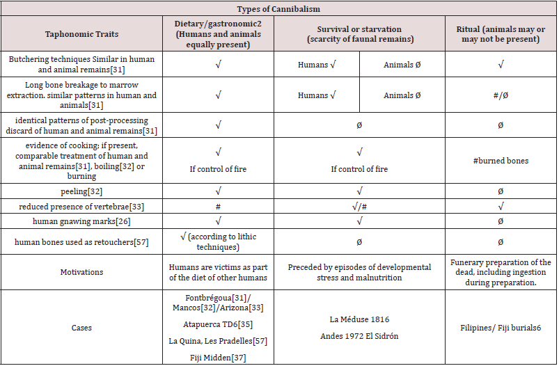
Dietary cannibalism was first described by Villa et al. [31] based on the Neolithic human remains found in Fontbrégua (France). The authors set up the basis of bone surface taphonomic features, spatial distribution traits and contexts to identify dietary cannibalism and to distinguish it from ritual or survival cannibalism. Later studies by White [32], Turner and Turner [33], Fernández-Jalvo et al. [34,35], Defleur et al. [36], and Degusta [37] have added traits that characterize both paleoanthropological and modern anthropological cases of dietary cannibalism (re-named as gastronomic cannibalism by White, 1992). Basically dietary cannibalism is characterized by:
i. Similar butchering techniques in human and animal remains
in an assemblage and therefore the same types of cut, chop,
sawing and scraping marks are identified in both animals and
humans, always considering anatomical differences between
robust large mammals compared to smaller human skeletons.
ii. similar patterns of long bone breakage to facilitate marrow
extraction;
iii. identical patterns of post-processing discard of human and
animal remains;
iv. evidence of cooking; if present, such evidence should indicate
comparable treatment of human and animal remains. Traits
of surface modifications and contexts of the different types of
cannibalism are displayed in Table 1.
Villa et al. [31] also noted that the presence of cut marks on
human remains may not be an exclusive indication for cannibalism
and they could be associated with secondary burials. This includes
traces of funerary rites involving the handling of corpses without
consumption of human tissues and includes dismemberment
and defleshing of the body. However, bone breakage for marrow
extraction and the mode of bone disposal and indication of food
discard serve to set funerary apart from dietary cannibalism.
Survival cannibalism has hunger as a motivation and is a
response to protein shortages, with scarce or absent faunal remains
(Table 1). Several cases of this type of cannibalism have been
described in recent times from well-known historical catastrophes,
such as the wreck of the frigate Méduse in 1816 or the Andean air
crash in 1972. Prehistoric survival cannibalism has been cited by
Rosas et al. [38] in the Spanish site of El Sidrón, apparently preceded
by episodes of developmental stress and malnutrition combined
with rarity of faunal remains (Table 1 and Figure 1).
The earliest known ritual cannibalism based on religious beliefs
was the Aztec’s pre-Columbian religions, described and figured in
codices of the 16th century that display domestic activities and
scenes of cannibalism [39-41]. Whether cannibals used religious
beliefs as a way to get an easy meal is a subject to be studied [41].
Taphonomic studies of some cases of prehistoric cannibalism, such
as the Anasazi at Mancos [32] and Los Pueblos of Arizona [33], have
shown indications of dietary cannibalism. Furthermore, Turner
and Turner [33] found that these groups sharpened their teeth to
frighten and threaten their neighbors, perhaps a further indication
of their cannibalistic traditions. A better distinction between ritual
mortuary practices and victims discarded as food remains has been shown by Degusta [37] at Fiji. This author showed clear differences
between consumed individuals mixed with animals by Fijians
(Navatu midden), and mortuary practices (Navatu burials) where
the buried dead showed few signs of consumption during rituals
(Table 1): they were not as damaged as cannibalized individuals,
and animal remains were not mixed with the buried humans.
Human remains from prehistoric sites such as Fontbrégua, Gran
Dolina-TD6 and El Mirador -Atapuerca and Gough’s Cave have
all been revised [25, 42-44] from an interpretation as dietary/
gastronomic cannibalism to an explanation highlighting a more
ritualistic or funerary connotation.
Table 1: The identification of human cannibalism and the purpose of such action is based on taphonomic features recorded on the bone surfaces and the context of the sites. These traits indicate a dietary or survival purpose, other than ritual practices. √= PRESENT & FREQUENT; #=Reduced presence or almost absent; Ø =absent.
One of the most extensive Magdalenian human bone assemblages comes from Gough’s Cave, a limestone cave set in Cheddar Gorge (Somerset, UK). This bone assemblage was considered to be result of cannibalistic activities during the Upper Palaeolithic (14,700 cal BP according to Jacobi and Highman, 2009) where Homo sapiens fossil bones record abundant tool-induced modifications and intense breakage [26]. Previous analyses of these fossils were undertaken in the 1980’s when trampling marks were seen to mimic cut marks, and the cut marks were interpreted with caution [45]. The latter author considered that most of the cuts were in fact scratches largely due to natural damage (trampling) except for one fossil (an adult mandible) with cut marks in the concave interior (lingual aspect) of the symphysis. In a later paper, [46] reconsidered that surface scratches on the Gough’s bones were made by stone tools to dismember corpses...BUT... she did not support dietary/gastronomic cannibalism: “...if cannibalism is implied, its practice could not have been a ... necessity because plenty of food seems to have been available at the site throughout the period of occupation” [45]. No indication of artistic engraving on human remains was ever described by Cook. These marks were later interpreted by Andrews and Fernández-Jalvo [26] as result of tongue removal, highly similar to other cuts found on the symphysis of a horse mandible at this site and result of nutritional cannibalism.
In addition, the skulls (and faces) of Gough’s Cave where described to “ have a higher intensity of cut-marks than are present on non-human animals, but contrasting with this, two of the human skulls are almost complete” “These contradictory results (strong damage and completeness) may suggest that the skulls were carefully treated to preserve them complete, in contrast to the rest of the skeleton and other animals at the site “, something ignored by Bello et al. [25].
Materials and Methods
The entire Gough’s Cave collection of both humans and large mammals housed at the Natural History Museum was analyzed by Andrews and Fernández-Jalvo [26], the fossil distribution plans and pictures taken from systematic excavations at the site were studied, and the site as it is nowadays adapted to touristic tours was visited. We could also add observations from controlled experiments between 1998 and 2000 to reproduce some characteristic taphonomic traits made with tools of different raw materials on long bones bearing little meat, and human chewing as well. All our results were described and published in Andrews and Fernández- Jalvo [26]. One of us (YFJ) previously studied the fossil collection from Gran Dolina TD6-Atapuerca [35,47] the oldest [48] pure gastronomic cannibalistic case with which to compare traits with the Gough’s Cave fossil assemblage.
Considering only the type of cut marks, Fernández-Jalvo and
Andrews [21] analyzed the peculiar cuts found on the human
radius of Gough’s Cave (M54074) and compared them to other
atypical cuts from butchery kill sites and the cannibalistic case
from TD6 at Atapuerca. Microscopic images at high magnification,
resolution and depth of field are an important and highly useful
tool in the study of taphonomic modifications. Optical microscopy
is improving in resolution and magnification, as well as through
the application of computer programs to treat serial images taken
from different heights to improve depth of field. Some of these
microscopes use the same principles as confocal microscopy
using natural white light. Confocal technology with natural light
of 3D microscope, more specifically Leica DCM8, is built based on
the same patent equipment as Alicona. The same 3D microscopic
equipment to validate descriptions given by Bello et al. [25] in their
analysis of cut marks. Nonetheless, the use of computer-generated
serial images to obtain a single image can lead to shading that
natural light creates when the modification is deep and inclined.
This may lead to identifications of profiles that are not real, hiding
significant details and wrongly interpreting the nature of the cut.
The principles of electron microscopy further augment the
capacity of optical microscopes in observation and analyses of
samples, greatly improving magnification, resolution and depth
of field. Scanning electron microscopy (SEM) combines imaging
with composition detectors of electrons emitted by the sample
when struck by an electron beam. The main image detectors are
secondary electrons (SE-SEM) that observe the topography of the
specimen. Backscattered electron mode (BSE-SEM) gives a flatter
image, but it provides important information on the density and
composition of the sample; high mineral content (brighter) versus
high organic content (darker).
Discussion: Dietary vs. ritual cannibalism in Gough’s cave
The Gough’s Cave human radius (M54074): decoration?
Elaborate cuts are present on a human radius (M54074) from Gough’s Cave (Figure 1a and 1b). They were analyzed in detail by Andrews and Fernández-Jalvo [26] who recognized a peculiar distribution and shape of these cut marks in an anatomical area that did not have any muscle attachment and bear little meat. The authors concluded that each group of marks were made by a single stroke incising the tool on the bone surface from different angles as determined by the user’s hand supination (Figs. 1c, 1d); we concluded that the multiple marks were the result of progressive filleting of flesh along the shaft. These same marks, however, suggested to Bello et al. [25] that each individual mark was a mixed combination of several actions (i.e. scraping, sawing and single strokes as shown in their publication). The resulting line of zig-zag cuts on the human radius were compared by Bello et al. [25] to the marks on decorated bones from Magdalenian sites, concluding that the Gough’s Cave radius is the oldest artistic manifestation on a human bone.
We have re-analyzed these cuts (Figure 1a) to find any possible mistakes in our previous interpretations. The characteristic shape of individual cuts can be distinguished in the row of cuts of M54074. In order to facilitate recognition of the shape of each stroke and the superposition of marks, we have outlined each movement in Figure 1b Each movement with a different angle and hand supination is outlined in red or in blue. The red marks precede the blue cuts which are superimposed on them. The black arrows point to a distinct trait that repeats on the left side of the cuts outlined in ‘blue’. This lateral salient has also been observed on fossil bones from Atapuerca (Trinchera Norte site, Middle Pleistocene, Figure 1c and on experimental cuts made by one of us (YFJ) during butchery. The experimental cuts (Figures 1d and 1e) correspond to individual cut marks (single movements) made when filleting a leg of lamb (a tibia) using an unretouched flint flake. The experiment was part of an experimental protocol aimed at distinguishing cut marks made by different raw materials of tool for a study published by Fernández-Jalvo et al. [35] that predates the work we did on the Gough’s Cave bones. Notes recorded during the experiment mentioned that the experimenter inclined the tool incising the edge almost horizontally relative to the bone surface, to better remove the meat. These cuts recorded the lateral salient irregularities of the tool.
The zig-zag pattern is related to the relative position of the cut-maker’s hand supination and the orientation of the long bone (distal end forward for the blue marks or downwards for the red marks), so that the apparent, overall zig-zag shape is actually the mirror image of each pair of cuts (bright green square in Figure 1b). This mirror pattern can be recognized in the subsequent cuts along the apparent zig-zag edge of the human radius M54074. The result of reiterative cutting movements along the shaft when filleting, as shown in Fig. 1b, changing the orientation of the bone respect to the cut-maker, caused the apparent zig-zag shape. The cuts, both those outlined in blue and in red, record a characteristic repetitive pattern of the tool edges, although sometimes with slight differences (the cuts outlined in red do not record the feature pointed with a black arrow in Figure 1b). These differences could probably be caused by the cut-maker’s handedness and hand supination. We could also speculate that the lack of records of some irregularities of the stone tool edge could be caused if the radius was still attached to the arm when filleting and the movement could not be identical when the distal end was forward or downward.
Figure 1: the cut marks) result of handedness, hand-supination, irregularity of the stone tool edge and moving the bone while filleting.
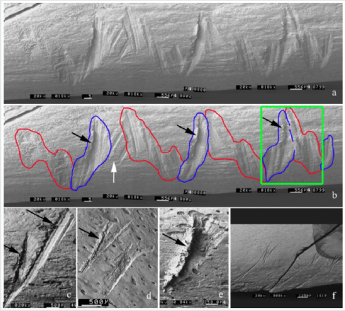
The experimental cuts on the butchered lamb tibia (Figures
1d and 1e) in the study published by Fernández-Jalvo et al. [35]
reproduce the shapes seen on the human radius from Gough’s Cave
(Figure 1b, outlined in blue) that are interpreted by Bello et al. [25]
as “scraping fan-shape” with a tool retouched on one side by single
or multiple movements. The striations outlined in red (Figure 1b)
have been interpreted by Bello et al.[25] as separate “to-and-fro
incisions”. In our opinion, the highly exhaustive and detailed 3D
microscopic inspection by Bello et al. [25] of each single individual
striation inside each of these cuts, has distorted the overall
interpretation of the actual cut shapes (Figure 1b).
Another case shown in Figure 1f displays three identical
cut-marks with distinct shapes that record stone tool edge
irregularities. Each cut was made in a single movement repeated
three times. These cut marks are identical to each other and were
made with identical hand supination by the cut- maker and the
bone orientation did not change when filleting [35,49].
A highly similar cut mark produced during butchery by one
of us (YFJ) has been observed using a similar equipment to that
used by Bello et al. [25] - a natural light confocal high-resolution
3D microscope (Leica DCM8). Comparing the striations made by us
when butchering (Figures1d, 1e and 2), and the cuts of the human
radius (M54074) analyzed in their Supplementary Information
(see engraving 1 and 65 Supplementary Information Bello et
al., [25], the profile shapes of the cut marks are almost identical.
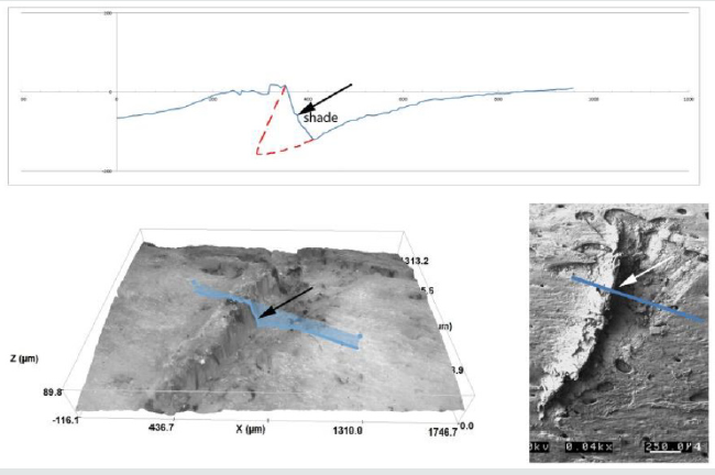
The profile of the cut obtained in the confocal high-resolution
equipment, however, is not real (Figure 2), for the lateral wall of the
cut appears vertical whereas in fact it slopes inwards. The shading
of the incident natural light on the sample distorts the 3D image
of Bello’s analysis and conceals traits that are characteristic and
typical of hand supination (Figure 2, broken red line) observed
using the scanning electron microscope (Figure1).
Figure 2: Image observed and analyzed with confocal technology with natural light of 3D Leica DCM8 (similar technique as Alicona). Top: profile of the cut mark in blue and correction of the shape of the cut in red. Bottom left: image of the bone surface showing the artefact caused by shades that natural light creates which are not well interpreted by the image treatment computer program. Bottom right: Scanning electron microscope microphotograph showing the incision made in a single movement during lamb butchery by one of us (YFJ).
The human radius M54074 of Gough’s Cave shows other cuts on
its surface that are clear slicing cut marks identified as butchery by
Andrews and Fernández-Jalvo [26] and by Bello et al. [25]as well.
These butchery cuts (Figure 1b, white arrow) predate some of the
zig-zag incisions (Figure 1b, white arrow) considered by Bello et
al., (2017) as decoration, while Andrews and Fernández-Jalvo [26]
considered them as filleting marks. In addition, the human radius
M54074 from Gough’s Cave was intensively broken to extract the
marrow after cutting. Intense breakage was also mentioned by
Bello et al. [25], and the question that rises is why decorate a bone
in between purely gastronomic activities shown by butchery cut
marks and marrow extraction? Besides cut marks and marrow
extraction, some Gough’s human bones were intensely chewed by
humans for nutrient exploitation [26,30]. Similarly, in Atapuerca TD6 and Mirador, chewing marks of other humans have also been
observed in the bones of the victims and animal bones [49-51]
indicating, initially, at least, pure gastronomic purposes.
The breakage of the human radius to obtain the marrow
(a purely gastronomic act) is not observed on the decorated
animal bones from Gough’s Cave which were handled and used
as indicated by the shiny patina that cover these specimens. The
decorative bones from Gough’s Cave include engraving and “bâtons
percés” on animal bones, but none of these decorated bones have
the complexity of actions as described on the human radius by Bello
et al. [25]. On the contrary, they have deep incisions and piercing
controlled by the cut-maker (Figure 2 in Bello et al., [25]) and are
not as superficial as those observed on the human radius.
The skull cups from Gough’s Cave
The purposeful component of the cannibalistic ritual at the site
has been linked to a complex mortuary ritual context reinforced by
the presence of complete skull calvaria which were interpreted as
“skull cups” indicating ritual purposes [30]. Skull caps have been
found in sites where cannibalism was practiced by Homo sapiens,
such as Herxheim [52], L’Adouste [53], Mirador [50] or Fiji [37]
where taphonomic traits indicated different types of cannibalism
related to meat consumption. Dietary cannibalism defined by Villa
et al., [31] at Fontbrégua yielded relatively complete skulls that were
suggested to be a kind of special treatment. Later, other authors
suggested that the isolated presence of these so-called “skull cups”
in Fontbrégua showed that the Fontbrégua assemblage could be
interpreted as ritual cannibalism [42]. This ritual motivation at
Fontbrégua, suggested by Bahn and other authors [53], is contrary
to the view of Villa et al. [54] who responded to Bahn that both
humans and animals were processed identically and, therefore, if
any ritual treatment is considered for human bones, animal bones
then would be expected to have had similar ritual treatments. This
interpretation was also mentioned in the Gough’s Cave paper, where
together with complete calvaria, animals and humans are mixed
together as discard food and have identical butchery techniques to
extract the skin, the meat, the viscera and the marrow [26].
An alternative hypothesis is based on the natural breakage of
human skulls, that preserves the calvaria complete (Figure 3). Homo
sapiens could see the practical bowl-like shape for domestic use
and perhaps tried to imitate natural skull breakage processing the
skulls to preserve them complete. This needed special skill, delicate
and careful treatment to prevent the calvaria accidentally breaking.
If preparation of skulls was not for manufacture of a temporary
utility but were made as a ritual treatment, it is hard to explain why
the skulls were abandoned with other body parts of these corpses
and mixed with butchered animal bones representing food refuse.
Figure 3: Different human calvaria naturally broken as a bowl (top and middle raws) compared to cannibalistic contexts where Homo sapiens was involved (bottom raw). Top: Homo erectus (Pekin Man, ©Wikipedia) Zhoukoudien skull calvaria. Middle raw: Neanderthal skulls from Engis (©Wikipedia) and Spy (Belgium, Courtesy of P.Semal) and Neander (Germany, ©Wikipedia). Bottom raw: skulls from Frontbrégua (France, Courtesy of P.Villa), Gough’s Cave (UK, © Natural History Museum) and El Mirador-Atapuerca (Spain, Courtesy of J.M.Vergés) all of them cannibalized by Homo sapiens.
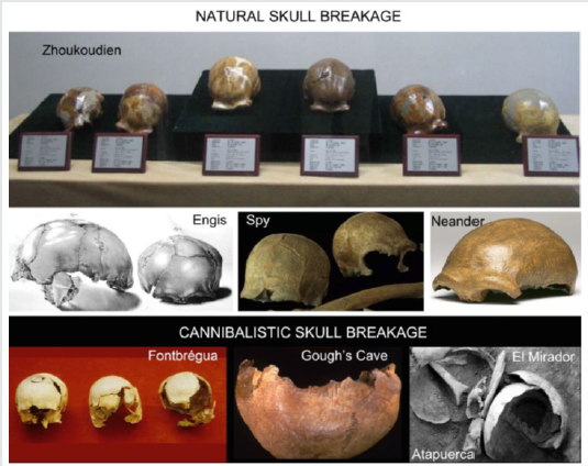
A recent paper by Marginedas et al. [44] found standardized
patterns of scalp removal with repetitive pattern and delicate
preparation on these calvaria. This pattern may reflect the
preparation of skulls to preserve them complete. The procedure to
remove skin, scalp and attached muscles are thus, not necessarily
a ritual treatment, but detachment of muscle insertions reflecting
anatomical dissection.
The presence of these complete calvaria in Gough’s Cave is
then contradictory to the idea of the site as a cannibalistic context,
with a high intensity of breakage and nutrient extraction in both
postcranial and cranial anatomical elements. Similar intensity of
butchery and breakage of hominin and animal skeletons noted in
Gough’s Cave has also been observed in Atapuerca-Gran Dolina TD6
[35], with the only exception of complete calvaria at Gough’s Cave.
None of the skull fragments from Atapuerca-TD6 are larger than
few centimeters. Especially remarkable in the cranial skeletons
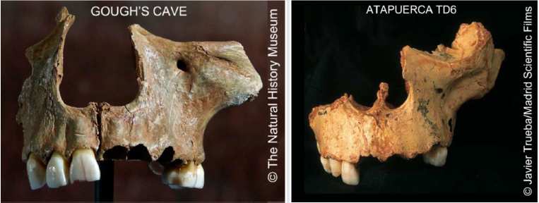
of Gough’s Cave and Atapuerca-TD6 are the faces, as part of the
most recognizable cranial skeleton of the human anatomy. Faces
appear in Gough’s Cave as intensively broken, with an identical
configuration, like those found in TD6 (Figure 4) as a result of
similar intense breakage and meat removal [55].
Figure 4: Faces of Gough’s Cave (top) compared to faces of Atapuerca TD6 (bottom). Inconsistent with the apparent care preparing skull cups is the intense breakage observed on the faces in Gough’s Cave.
Final Remarks
Mimicking cut marks from different processes are frequent
in taphonomy and the case of the human radius of Gough’s Cave
is a good example. Cut marks on the human radius from this
site have highly similar, if not identical shapes, to those we have
experimentally produced when meat was removed using individual
movements due to hand supination and irregularities of the tool
edge.
There are marked differences between the cut marks used
for bone decoration as found in other decorated specimens from
Gough’s Cave site, such as ‘battons percés’ which are characterized
by individual and deep cuts, and those on the human radius.
Similarities found by Bello et al., [25] with Magdalenian decorative
bones (based on an apparent overall zig-zag aspect of the cuts [56])
are the result of mimicking marks from different processes, i.e.
equifinality, so frequent in taphonomy.
The unusual shape of cuts on the human radius results from
the confluence of several factors, such as the type of bone (a
radius which bears less meat than other anatomical elements),
lateral irregularities of the edge shape of the tool (bearing salient
irregularities that marked the bone when cutting), and the cutmaker,
who inclined the stone tool, maybe due to lack of skill, and did
not make a straight cut. Given that each set of striations outlined in
Figure 1b is superimposed on others, both groups of marks should
be equally the result of subsequent cutting movements during
a butchery process, using the same tool but with different hand
supination by the cut-maker, maybe changing the orientation of the
bone respect to the butcher. The overall zig-zag shape of the cuts is
actually the result of handedness, hand-supination, irregularity of
the stone tool edge and moving the bone while filleting.
All these indications suggest that the complex cuts repeated
along the length of the Gough’s human radius were the result of
butchery and filleting when removing meat, without decorative
intention.
The other factor considered to be the outcome of ritual
treatment is the complete calvaria named as skull cups. Skull cups
appear in cannibalistic cases practiced by Homo sapiens. Whether
these are indication of rituals or just a domestic and useful
modification of the skull, imitating natural breakage of skulls to
obtain a recipient or bowl shape, should be investigated in depth. Most cases where these skull cups have been found are in a context
of intense nutritional exploitation of human bodies that, with
the exception of the skulls, have all traits of dietary/gastronomic
cannibalism. Notably, the general context of Gough’s Cave is one of
intense nutritional exploitation, very similar to Gran Dolina-TD6.
Both of them are characterized by intense and identical breakage,
even of human faces, comparable treatment of humans and animals
to obtain nutrients, and the mixing of the human remains with
animal bones as food discard. Apparent ritualistic treatment of the
skulls could more likely be due to transformation of the shape of the
skull to obtain a bowl shape for practical domestic use rather than
ritual motivations.
The most parsimonious hypothesis for Gough’s Cave then
is that the human radius, like the rest of the human skeletal
elements, including skulls and faces, were intensively butchered in
cannibalistic acts and deposited in a context in which human bones
were with other animals intensively butchered
Acknowledgements
We are grateful to the technicians of the Electron Microscopy Unit of the Natural History Museum 335 and the Laboratory of Non-destructive Techniques of the Museo Nacional de Ciencias Naturales for their professional work. We thank Liora K. Horwitz for comments on the manuscript and to Crystal Jones for inviting the senior author to contribute to the Journal of Anthropological and Archaeological Sciences. Research described in this paper has been partially funded by the Spanish National Program of Research and Competitivity (project CGL2016-79334-P).
References
- Ambrose SH (2001) Paleolithic technology and human evolution. Science 291: 1748-1753.
- Plummer T (2004) Flaked stones and old bones: Biological and cultural evolution at the dawn of technology. Yearbook of Physical Anthropology 47: 118–164.
- Gowlett JAJ (2009) Artefacts of apes, humans, and other: Towards comparative assessment andanalysis. Journal of Human Evolution 5: 401-410.
- Whiten A, Schick K, Toth N (2009) The evolution and cultural transmission of percussive technology: Integrating evidence from paleoanthopology and primatology. Journal of Human Evolution 57: 420-435.
- Nonaka T, Bril B, Rein R (2010) How do stone knappers predict and control the outcome of flaking? Implications for understanding early stone tool technology. Journal of Human Evolution 59: 155-167.
- Andrews P, Cook J (1985) Natural modifications to bones in a temperate setting. Man 20: 675-691.
- Behrensmeyer AK, Gordon KD, Yanagi GT (1986) Trampling as a cause of bone surface damage and pseudo cutmarks. Nature 319: 768-771.
- Olsen A, Shipman P (1988) Surface modification on bone: trampling vs. butchery. Journal of Archaeological Science 15: 535-553.
- Shale Y, El Zaatari S, White TD (2017) Hominid butchers and biting crocodiles in the African Plio-Pleistocene. Proceedings of National Academy of Science.
- Domínguez-Rodrigo M, Baquedano E (2018) Distinguishing butchery cut marks from crocodile bite marks through machine learning methods. Nature. Scientific Reports 8: 5786.
- Bromage TG, Boyde A (1984) Microscopic criteria for the determination of directionality of cutmarks on bone. American Journal of Physical Anthropology 65: 359–366.
- Bromage TG, Bermudez de Castro JM, Fernández-Jalvo Y (1991) The SEM in taphonomic researchand its application to studies of curmarks generally and the determination of handedness specifically. Anthropologie 29: 163-169.
- Bello SM, Parfitt SA, Stringer CB (2009) Quantitative micromorphological analyses of cut marks produced by ancient and modern handaxes. Journal of Archaeological Science 36: 1869-1880.
- de Juana S, Galán AB, Domínguez-Rodrigo M (2010) Taphonomic identification of cut marks made with lithic handaxes: An experimental study. Journal of Archaeological Science 37: 1841–1850.
- Yravedra J, Domínguez-Rodrigo M, Santonja M, Pérez-González A, Panera J, et al. (2010) Cut marks on the Middle Pleistocene elephant carcass of Áridos 2 (Madrid, Spain). Journal of Archaeological Science 37: 2469-2476.
- MacPherron SP, Alemseged Z, Marean CW, Wynn JG, Reed D, et al. (2010) Evidence for stone-tool-assisted consumption of animal tissues before 3.39 million years ago at Dikika, Ethiopia. Nature 466: 857–860.
- Pickering T, White T, Toth N (2000) Brief Communication: Cutmarks on a Plio-Pleistocene Hominid From Sterkfontein, South Africa. Journal of Physical Anthropology 111: 579-584.
- Domínguez-Rodrigo M, Pickering TR, Bunn H (2010) Configurational approach to identifying the earliest hominin butchers. Proceedings of National Academy of Science 107: 20929-20934.
- Hanon R, Péan S, Prat S (2018) Reassessment of Anthropic Modifications on the Early Pleistocene Hominin Specimen Stw53 (Sterkfontein, South Africa). Bulletins et Mémoires de la Société d’Anthropologie de Paris 30: 49-58.
- Bello SM, Soligo C (2008) A new method for the quantitative analysis of cutmark micromorphology. Journal of Archaeological Science 35: 1542-1552.
- Fernández-Jalvo Y, Andrews P (2016) Atlas of Taphonomic Identifications: 1001+ images of fossil and recent mammal bone modification. Dordrecht: Springer.
- Harris et al. (2017) The trajectory of bone surface modification studies in paleoanthropology and a new Bayesian solution to the identification controversy. Journal of Human Evolution 110: 69-81.
- Domínguez-Rodrig M, Saladié P, Cáceres I, Hughet R, Yravedra J, et al. (2017) Use and abuse of cut mark analyses: The Rorschach effect. Journal of Archaeological Science 86: 14-23.
- Cifuentes-Alcobendas G, Domínguez-Rodrigo M (2019) Deep learning and taphonomy: High accuracy in the classification of cut marks made on fleshed and defleshed bones usingconvolutional neural networks. Nature Scientific Reports 9: 18933.
- Bello SM, Wallduck R, Parfitt SA, Stringer CB (2017) An Upper Palaeolithic engraved human bone associated with ritualistic cannibalism. PLoS ONE 12: e0182127.
- Andrews P, Fernández-Jalvo Y (2003) Cannibalism in Britain: Taphonomy of the Creswellian(Pleistocene) faunal and human remains from Gough's Cave (Somerset, England). Bulletin of the Natural History Museum, London. Geology 58: 59–81.
- Breuil L’Abbé H, Obermaier H (1909) Crânes Paléolithiques façonnés en coupes. L’Anthropologie XX: 523–530.
- Le Mort F, Gambier D (1991) Cutmarks and breakage on the human bones from Le Placard (France). An example of special mortuary practice during the Upper Palaeolithic Anthropology 29: 189–194.
- Buisson D, Gambier D (1991) Façonnage et gravure sur des os humains d’Isturitz (Pyrenées-Atlantiques). Bulletin de la Société préhistorique française 88: 174–177.
- Bello SM, Parfitt SA, Saladié P, Cáceres I, Rodríguez-Hidalgo A (2015) Upper Palaeolithic ritualistic cannibalism: Gough's Cave (Somerset, UK) from head to toe. Journal of Human Evolution 82: 170-189.
- Villa P, Bouville C, Courtin J, Helmer D, Mahieu E, et al. (1986) Cannibalism in the Neolithic. Science 233: 431–437.
- White TD (1992) Prehistoric Cannibalism at Mancos 5MTUMR-2346. Princeton University Press, NJ, USA.
- Turner CG II, Turner JA (1999) Man Corn: Cannibalism and Violence in the Prehistoric American Southwest. University of Utah Press, USA.
- Fernández-Jalvo Y, Díez JC, Bermúdez de Castro JM, Carbonell E, Arsuaga JL (1996) Evidence of early cannibalism. Science 271: 277-278.
- Fernández-Jalvo Y, Díez, JC, Rosell J, Cáceres I (1999) Human Cannibalism in the Early Pleistocene of Europe (Gran Dolina, Sierra de Atapuerca, Burgos, Spain). Journal of Human Evolution 37: 591-622.
- Defleur A, White T, Valensi P, Slimak L, Crégut-Bonnoure E (1999) Neanderthal cannibalism at Moula-Guercy, Ardèche, France. Science 286: 128–131.
- Degusta D (1999) Fijian cannibalism: Osteological evidence from Navatu. American Journal of Physical Anthropology 110: 215–241.
- Rosas A, Martínez-Maza C, Bastir M, García-Tabernero A, Lalueza-Fox C, Huguet R, et al. (2006) Paleobiology and comparative morphology of a late Neandertal sample from El Sidron, Asturias, Spain. Proceedings of National Academy of Science 103: 19266-19271.
- Staden Jv (1558) Waerachtige Historie en beschrijvinge eens Landts in America gheleghen, wiens inwoonders Wilt, Naect, seer godtloos ende wreede Menschen eeters zijn. Antwerpen. Amsterdam.
- Batalla JJ (1999) El Códice Tudela o Códice del Museo de América y el Grupo Magliabechiano. Unpublished PhDThesis. Universidad Complutense of Madrid.
- Wade L (2018) Feeding the Gods. Science 360(6395): 1288-1292.
- Bahn PG (1990) Eating people is wrong. Nature 348: 395.
- Carbonell E, Cáceres I, Lozano M, Saladié P, Rosell J, Lorenzo C, et al. (2010) Cultural cannibalism as a paleoeconomic system in the European Lower Pleistocene. Current Anthropology 51: 539–549.
- Marginedas F, Rodríguez-Hidalgo A, Soto M, Bello SM, Cáceres I, et al. (2020) Making skull cups: butchering traces on cannibalised human skulls from five European archaeological sites. Journal of Archaeological Science 114: 105076.
- Cook J (1986) Marked human bones from Gough’s Cave Somerset. 1986. Proceedings of the University of Bristol Spelaeological Society 17: 275-285.
- Cook J (1991) Preliminary report on marked human bones from the 1986-1987 excavations at Gough's Cave, Somerset, England. Anthropologie (Brno) 29(3): 181-187.
- Díez JC, Fernández-Jalvo Y, Rosell J, Cáceres I (1999) The site formation (Aurora Stratum, Gran Dolina, Sierra de Atapuerca, Burgos, Spain). Journal of Human Evolution 37: 623–652.
- Pares JM, Arnold L, Duval M, Demuro M, Pérez-González, Bermúdez de Castro JM, et al. (2013) Reassessing the age of Atapuerca-TD6 (Spain): New paleomagentic results. Journal of Archaeological Science 40: 4586-4595.
- Fernández-Jalvo Y, Andrews P (2011) When human chew bones. Journal of Human Evolution 60: 117-123.
- Cáceres I, Lozano M, Saladié P (2007) Evidence for Bronze Age Cannibalism in El Mirador Cave (Sierra de Atapuerca, Burgos, Spain). American Journal of Physical Anthropology133: 899-917.
- Saladié P, Rodríguez-Hidalgo A, Díez C, Martín-Rodríguez M, Carbonell E (2013) Range of bone modifications by human chewing. Journal of Human Evolution 40: 380-397.
- Boulestin B, Zeeb-Lanz A, Jeunesse C, Haack F, Arbogast RM, Denaire A (2009) Mass cannibalism in the linear pottery cultures at Herxheim (Palatinate, Germany). Antiquity 83: 968–982.
- Mafart B, Baroni I, Onoratini G (2004) Les restes humains de la Grotte de L'Adaouste du Neolithique Ancien final (Bouches du Rhone, France): Cannibalisme rituel or funerarire? British Archaeological Repoorts. International Series S 1303: 289–294.
- Villa P (1991) Cannibalism in the Neolithic. Nature 351: 613-614.
- Jacobi RM, Higham TFG (2009) The early Lateglacial re-colonization of Britain: New radiocarbon evidence from Gough's Cave, southwest England. Quaternary Sccience Review 28: 1895-1913.
- Lucas C (2016) L’art géométrique de Duruthy (Sorde-l’Abbaye, Landes, France): Du Magdalénien moyen au Magdalénien supé Actes du colloque « L'art au quotidien - Objets ornés du Paléolithique supérieur », Les Eyzies-de-Tayac, 16-20 juin 2014 PALEO, numéro spécial p.1-13.
- Verna C, d'Errico F (2011) The earliest evidence for the use of human bone as a tool. Jornal of Human Evolution 60: 145-157.

Top Editors
-

Mark E Smith
Bio chemistry
University of Texas Medical Branch, USA -

Lawrence A Presley
Department of Criminal Justice
Liberty University, USA -

Thomas W Miller
Department of Psychiatry
University of Kentucky, USA -

Gjumrakch Aliev
Department of Medicine
Gally International Biomedical Research & Consulting LLC, USA -

Christopher Bryant
Department of Urbanisation and Agricultural
Montreal university, USA -

Robert William Frare
Oral & Maxillofacial Pathology
New York University, USA -

Rudolph Modesto Navari
Gastroenterology and Hepatology
University of Alabama, UK -

Andrew Hague
Department of Medicine
Universities of Bradford, UK -

George Gregory Buttigieg
Maltese College of Obstetrics and Gynaecology, Europe -

Chen-Hsiung Yeh
Oncology
Circulogene Theranostics, England -
.png)
Emilio Bucio-Carrillo
Radiation Chemistry
National University of Mexico, USA -
.jpg)
Casey J Grenier
Analytical Chemistry
Wentworth Institute of Technology, USA -
Hany Atalah
Minimally Invasive Surgery
Mercer University school of Medicine, USA -

Abu-Hussein Muhamad
Pediatric Dentistry
University of Athens , Greece

The annual scholar awards from Lupine Publishers honor a selected number Read More...


