Lupine Publishers Group
Lupine Publishers
Menu
Research Article(ISSN: 2770-5447) 
Acute Pulmonary Thromboembolism with Patent Foramen Ovale and Right Lower Limb Arterial Gangrene: A Case Of Paradoxical Embolism Volume 3 - Issue 5
Aamir Shafi*, Wani Abdul Ahad and Jawhar ul Islam
- Department of Medicine SKIMS Medical College Bemina Srinagar, India
Received:February 11, 2022; Published:February 28, 2022
Corresponding author:Aamir Shafi Department of General Medicine, SKIMS Medical College and Hospital, Bemina Srinagar, Jammu and Kashmir, India
DOI: 10.32474/ACR.2022.03.000175
Abstract
Paradoxical Embolism with acute arterial ischemia is an uncommon but known entity in medical literature. However, existence of concomitant pulmonary thromboembolism with a patent foramen ovale presenting as limb arterial gangrene has not been reported in literature to the best of our knowledge and search. we present a case of right lower limb arterial gangrene in an elderly female who had concomitant Pulmonary thromboembolism with a patent foramen ovale. The limb gangrene was attributed secondary to paradoxical embolism due to patent foramen ovale. The patient was managed with limb amputation and long term oral anticoagulation.
Keywords: Paradoxical Embolism; Gangrene; Patent Foramen Ovale
Case
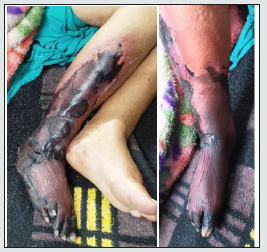
A 65-year-old female from a tribal area of Kashmir with no existing co morbidities presented to our emergency department with progressive shortness of breath and blackish discoloration of right lower limb of 3 weeks duration. It was preceded by dull aching right lower limb pain which would worsen with walking few steps. There was no such similar history in the past or any use of OCPs or lipid disorders. she had no pregnancy losses during her reproductive age. On examination she was tachypneic with respiratory rate of 26 breaths per minute and hypoxia with an oxygen saturation of 88% while breathing ambient air. There was no hypotension or focal neurological deficit. cardiac auscultation did not reveal any heave or murmur. Examination of right lower limb revealed gross blackish limb distal to knee joint with overlying skin necrosis (Figure 1). Baseline investigations including chest x ray and ECG were normal. Popliteal, dorsalis pedis and posterior tibial pulses were absent .Color Doppler study of lower limb revealed total occlusion of popliteal artery with no distal flow. In view of unexplained hypoxemia and raised D dimer she was subjected to CT pulmonary angiography which revealed an eccentric thrombus in right pulmonary artery (Figure 2). Thrombophilia profile did not reveal any evidence of hypercoagulable state. Keeping in view PTE with concomitant arterial thrombosis, a possibility of paradoxical embolism was made. She underwent Transthoracic echocardiography which revealed Patent foramen ovale (Figure 3). She was subjected to above knee amputation and referred to cardiology centre for closure of PFO for secondary prevention and discharged on long term oral anticoagulation.
Figure 1:Gangrenous right leg.

Discussion and review of literature
Paradoxical embolism is an uncommon cause for acute arterial occlusion. Paradoxical Embolism (PDE) occurs when a thrombus crosses an intracardiac defect into the systemic circulation [1,2]. Patients may present with symptoms based on the site of the resultant embolization. These sites can include the brain, heart, gastrointestinal tract, or extremities(reference case) [3]. Patients may present with a cerebrovascular event, chest pain,, cold extremity, or mesenteric ischemia. The embolus is usually a blood clot but may be a fat particle, air, amniotic fluid, or tumor [1,3]. The management of paradoxical embolism is medical and/or surgery, depending on the location of the embolus.
Etiology
Patent foramen ovale (PFO): is a left to right shunt that
occurs between the septum primum and septum secundum. The
magnitude of the right to left shunt may be associated with an
increased risk of cryptogenic stroke [4]. Valsalva maneuvers such
as coughing, squatting, or defecating can transiently increase
right atrial pressure leading to a transient shunt reversal and
the transfer of potential thrombi into the systemic circulation.
Clinically significant PFOs result in adverse consequences by two
mechanisms:
a) Serving as a conduit for paradoxical embolization from venous
side to systemic circulation
b) Because of their tunnel like structrure and propensity for
the stagnant flow ,may serve as nidus for in situ thrombus
formation.
The risk for a cryptogenic stroke in pts with PFO increases with larger defects and the presence of interatrial aneurysm (perhaps because of increase in situ thrombus formation in the aneurysmal tissue or because PFOs associated with an interatrial septal aneurysm tend to be larger).Despite prior reports concerning paradoxical embolism through PFO, this phenomenons magnitude as a risk factor for stroke or distal arterial embolism ( in our case) remains unidentified because deep vein thrombosis is infrequently detected in such patients. In one study pelvic vein thrombi were found more frequently in young patients with cryptogenic stroke than with those with known cause of stroke. This finding may provide the source of venous thrombi ,mainly when VTE is not identified initially [5,6]. A PFO is usually detected by Transthoracic echocardiography(TTE) or transcranial Doppler. Transesophageal echocardiography (TEE) is most sensitive test mainly when performed with contrast media injected during cough or valsalva maneuver [7]. In adults treatment for PFO is mainly indicated in a patient with cryptogenic stroke and other arterial embolic events. Medical therapy mainly includes antithrombotic medications (aspirin) especially in those with stroke due to embolism. Anticoagulation is indicated if stroke is associated with vein thrombosis. Atrial septal defects (ASD): are congenital defects that vary in size and location. ASDs lead to a left to right shunt as well as a fixed split S2 on cardiac exam. A transient reversal of blood flow can reverse the shunt. ASDs are associated with a paradoxical embolism in up to 14% of patients [5]. Ventricular septal defects: commonly result in left to right shunts, however certain conditions that increase right atrial pressure like Eisenmenger syndrome can reverse the shunt, allowing for paradoxical embolism. Pulmonary arteriovenous malformations are usually hereditary and are a pathological connection between the pulmonary arteries to the pulmonary veins returning to the left atrium. This leads to a permanent right to left shunt. Patients with a history of hereditary hemorrhagic telangiectasia are at increased risk for PAVM and subsequent paradoxical embolism[8].
Epidemiology
A PFO may be found in up to 30% of the population and studies have suggested that the annual risk of cryptogenic and recurrent strokes in patients with a PFO is 0.1% and 1% respectively [1,4]. ASDs are 2 to 3 times more common in females and are responsible for over 30% of congenital heart defects in adults [1]. Paradoxical embolism should be suspected in all patients with an ischemic stroke or other arterial thrombosis without an identifiable cause.
Pathophysiology
A paradoxical embolism can occur when a thrombus in the deep venous circulation embolizes through an intracardiac shunt or pulmonary artery venous malformation (PAVM) into the systemic circulation [1]. Deep venous thrombosis is a risk factor for paradoxical embolism. Studies have shown that cryptogenic stroke is 5 times more likely with pelvic vein thrombosis [9,10]. The pathophysiological mechanism of a paradoxical embolism does vary. In the setting of a PFO, any permanent increase in right-sided cardiac pressures can increase the risk of a paradoxical embolism [1].
Approach to patient
Paradoxical Embolism can be difficult to diagnose and have an
insidious onset. Physicians should strongly suspect paradoxical
embolism in patients with an embolic event with a non-identifiable
source, such as atrial fibrillation, and when a concomitant
intracardiac shunt or PAVM is known or suspected [1]. A thorough
history and physical examination are paramount. Depending on
the organ affected, the symptoms may vary.The most important
issues which should be addressed are [1-3].
a) Evaluation of factors that lead to the event (ie: coughing or
straining)
b) Screening for HTN, DVT, CVD, DM, hypercholesterolemia, atrial
fibrillation, stroke, and syncope.
c) History of migraines (migraines occurs in up to 50% of patients
with an intracardiac shunt).
d) History of congenital heart disease, structural heart disease, or
patent foramen ovale.
e) Family history
f) Social history, tobacco use.
Physical Examination Findings:
a. Physicians should look for signs of congenital heart defects
such as right ventricular hypertrophy, digital clubbing, or fixed
S2 splitting.
b. Full neurological evaluation (i.e.: speech or visual abnormality,
unilateral weakness, seizures, and swallowing difficulties).
c. Peripheral pulse evaluation and assessment for limb ischemia
as in our reference case (i.e.: extremity involved may present
with a sudden onset, be cold, pulseless, and painful) [3,4].
Evaluation
The diagnosis of paradoxical embolism is one of exclusion.
Other causes for the patient’s signs and symptoms should be
considered first. The evaluation should be as under [1-4,11].
a. ECG to assess for arrhythmia or atrial fibrillation.
b. Transthoracic echocardiogram (TTE) with color-flow Doppler
to evaluate for intracardiac shunts, cardiac myxomas, and
thrombus formations. Agitated saline or contrast can also
be injected during TTE to help visualize and diagnose an
intracardiac shunt. However, to accurately diagnose a PFO, the
saline or contrast needs to be injected at the end of a Valsalva
maneuver where the left of right shunt transiently reverses.
TTE can also be used to evaluate for the presence of aortic
plaques in the ascending aorta.
c. Transcranial Doppler sonography (TCD) to detect any shunt,
including PAVM. TCD is non-invasive and can be done at the
bedside by injecting agitated contrast into a peripheral line
and looking for microemboli in the middle cerebral artery..
d. Coagulation studies, including baseline coagulogram and
specific thrombophilic profile keeping in view clinical scenario
of the patient.
e. D-Dimer levels if pulmonary embolism is suspected
f. Arterial blood gas will help determine the oxygenation and
ventilation status.
g. Ultrasound and colour Doppler to evaluate for a DVT/arterial
occlusion.
h. CT angiography scan of the chest to assess for a pulmonary
embolus.
i. Noncontrast CT scan of the brain to assess for cerebrovascular
infarcts.
j. CT angiogram or MRI for acute occlusion of the extremity,
renal arteries , or bowel ischemia.
k. Ancillary blood chemistries and urinalysis as indicated to
assess for other metabolic causes.
Treatment/Management
The treatment of paradoxical embolism is based on both
medical and surgical approaches. The treatment pathways are
divided into three general approaches [1,3,4].
a. Elimination of the pathway allowing embolization (through
surgical or percutaneous approach)
b. Medical treatment to prevent recurrent episodes of venous
thrombosis.
c. Combination of medical and surgical approaches.
The choice of either and its specific plan is dependent on the risk of stroke recurrence, the lifelong benefit/risk ratio between antithrombotic therapy and surgery, as well as the cost of each intervention. The surgical approach includes occlusion of intracardiac shunts and PAVMs. Medical therapy is comprised of antithrombotic therapy, which includes aspirin, or clopidogrel as monotherapy or taken in combination with warfarin for the prevention of thrombotic events. The initial treatment is always anticoagulation. If the patient has an intracardiac communication, it may be closed percutaneously or with open-heart surgery. Today, many types of devices are available to close cardiac shunts percutaneously with minimal morbidity. Thrombolysis is often used in acute cases where the patient is hemodynamically unstable. Both DVT and pulmonary embolus need long term anticoagulation treatment. Some patients may need to be on antiplatelet therapy for life.
Prognosis
The prognosis after paradoxical embolism depends on the organ affected and the extent of the injury. Patients with CNS events usually tend to fare worse. Those who suffer mesenteric ischemia, or a cold leg may be salvaged with surgery(our reference case). On the other hand, renal infarction usually never recovers.
Conclusion
Paradoxical embolism with acute limb ischemia is a known entity in literature but our case with a PTE and PFO leading to arterial gangrene is a first case of its kind and after searching extensive literature we could not find replica of our reference case. To the best of our knowledge and review of literature we believe our patient had a pelvic vein thrombus which got embolized concomitantly into pulmonary artery and via PFO into right popliteal artery leading to pulmonary embolism and right lower limb gangrene respectively.
References
- Windecker S, Stortecky S, Meier B (2014) Paradoxical embolism. J Am Coll Cardiol 64(4): 403-415.
- Saremi F, Emmanuel N, Wu PF, Wu PF, Ihde L, et al. (2014) Paradoxical embolism: role of imaging in diagnosis and treatment planning. Radiographics 34(6): 1571-1592.
- Geng J, Tian HY, Zhang YM, He S, Ma Q (2017) Paradoxical embolism: A report of 2 cases. Medicine (Baltimore) 96(26): e7332.
- Maron BA, Shekar PS, Goldhaber SZ (2010)Paradoxical embolism. Circulation 122(19): 1968-1972.
- Bannan A, Shen R, Silvestry FE, Herrmann HC (2009) Characteristics of adult patients with atrial septal defects presenting with paradoxical embolism. Catheter Cardiovasc Interv 74(7): 1066-1069.
- Zoltowska DM, Thind G, Agrawal Y, Gupta V, Kalavakunta JK (2018) May-Thurner Syndrome as a Rare Cause of Paradoxical Embolism in a Patient with Patent Foramen Ovale. Case Rep Cardiol pp. 3625401.
- Smart D, Mitchell S, Wilmshurst P, Turner M, Banham N (2015) Joint position statement on persistent foramen ovale (PFO) and diving. South Pacific Underwater Medicine Society (SPUMS) and the United Kingdom Sports Diving Medical Committee (UKSDMC). Diving Hyperb Med 45(2): 129-131.
- Kjeldsen AD, Oxhøj H, Andersen PE, Green A, Vase P (2000) Prevalence of pulmonary arteriovenous malformations (PAVMs) and occurrence of neurological symptoms in patients with hereditary haemorrhagic telangiectasia (HHT). J Intern Med 248(3): 255-262.
- Osgood M, Budman E, Carandang R, Goddeau RP, Henninger N (2015) Prevalence of Pelvic Vein Pathology in Patients with Cryptogenic Stroke and Patent Foramen Ovale Undergoing MRV Pelvis. Cerebrovasc Dis 39(3-4): 216-223.
- Cramer SC, Rordorf G, Maki JH, Kramer LA, Grotta JC (2004) Increased pelvic vein thrombi in cryptogenic stroke: results of the Paradoxical Emboli from Large Veins in Ischemic Stroke (PELVIS) study. Stroke 35(1): 46-50.
- Karttunen V, Ventilä M, Ikäheimo M, Niemelä M, Hillbom M (2001) Ear oximetry: a noninvasive method for detection of patent foramen ovale: a study comparing dye dilution method and oximetry with contrast transesophageal echocardiography. Stroke 32(2): 448-453.

Top Editors
-

Mark E Smith
Bio chemistry
University of Texas Medical Branch, USA -

Lawrence A Presley
Department of Criminal Justice
Liberty University, USA -

Thomas W Miller
Department of Psychiatry
University of Kentucky, USA -

Gjumrakch Aliev
Department of Medicine
Gally International Biomedical Research & Consulting LLC, USA -

Christopher Bryant
Department of Urbanisation and Agricultural
Montreal university, USA -

Robert William Frare
Oral & Maxillofacial Pathology
New York University, USA -

Rudolph Modesto Navari
Gastroenterology and Hepatology
University of Alabama, UK -

Andrew Hague
Department of Medicine
Universities of Bradford, UK -

George Gregory Buttigieg
Maltese College of Obstetrics and Gynaecology, Europe -

Chen-Hsiung Yeh
Oncology
Circulogene Theranostics, England -
.png)
Emilio Bucio-Carrillo
Radiation Chemistry
National University of Mexico, USA -
.jpg)
Casey J Grenier
Analytical Chemistry
Wentworth Institute of Technology, USA -
Hany Atalah
Minimally Invasive Surgery
Mercer University school of Medicine, USA -

Abu-Hussein Muhamad
Pediatric Dentistry
University of Athens , Greece

The annual scholar awards from Lupine Publishers honor a selected number Read More...


